Figure 2. Paeoniflorin (PF) protects
human retinal pigment epithelium cells from H2O2-induced
cell death. A: Cytotoxicity of paeoniflorin to ARPE-19
cells were measured by 3-(4, 5-dimethylthiazol-2-yl)-2, 5
diphenyl tetrazolium bromide (MTT) assay. ARPE-19 cells were
treated with or without different concentrations of PF (5–200
μg/ml) for 24 h, and cell viability was assessed by an MTT
assay. The results are expressed as percentage of control, and
each value represents the mean±SEM of three independent
experiments (n=3 experiments). B: Cell viability of
ARPE-19 cells following H2O2 exposure were
measured by MTT assay. The cells were treated with or without
different concentrations of H2O2 (50–800
μM) for 24 h. Cell viability was measured by an MTT assay. The
results are expressed as percentage of control, and each value
represents the mean±SEM of three independent experiments (n=3
experiments, *p<0.05). C: Cell viability was measured
in ARPE-19 cells treated with 200 μM H2O2
or different concentrations of PF (50–200 μg/ml) by an MTT
assay. The results are expressed as percentage of control, and
each value represents the mean±SEM of three independent
experiments (n=3 experiments, *p<0.05). D: Cell
viability was measured using 4', 6-diamidino-2-phenylindole
(DAPI) staining. Strong fluorescent spots show apoptotic nuclei.
Figures were selected as representative data from three
independent experiments. Scale bar, 10 μm.
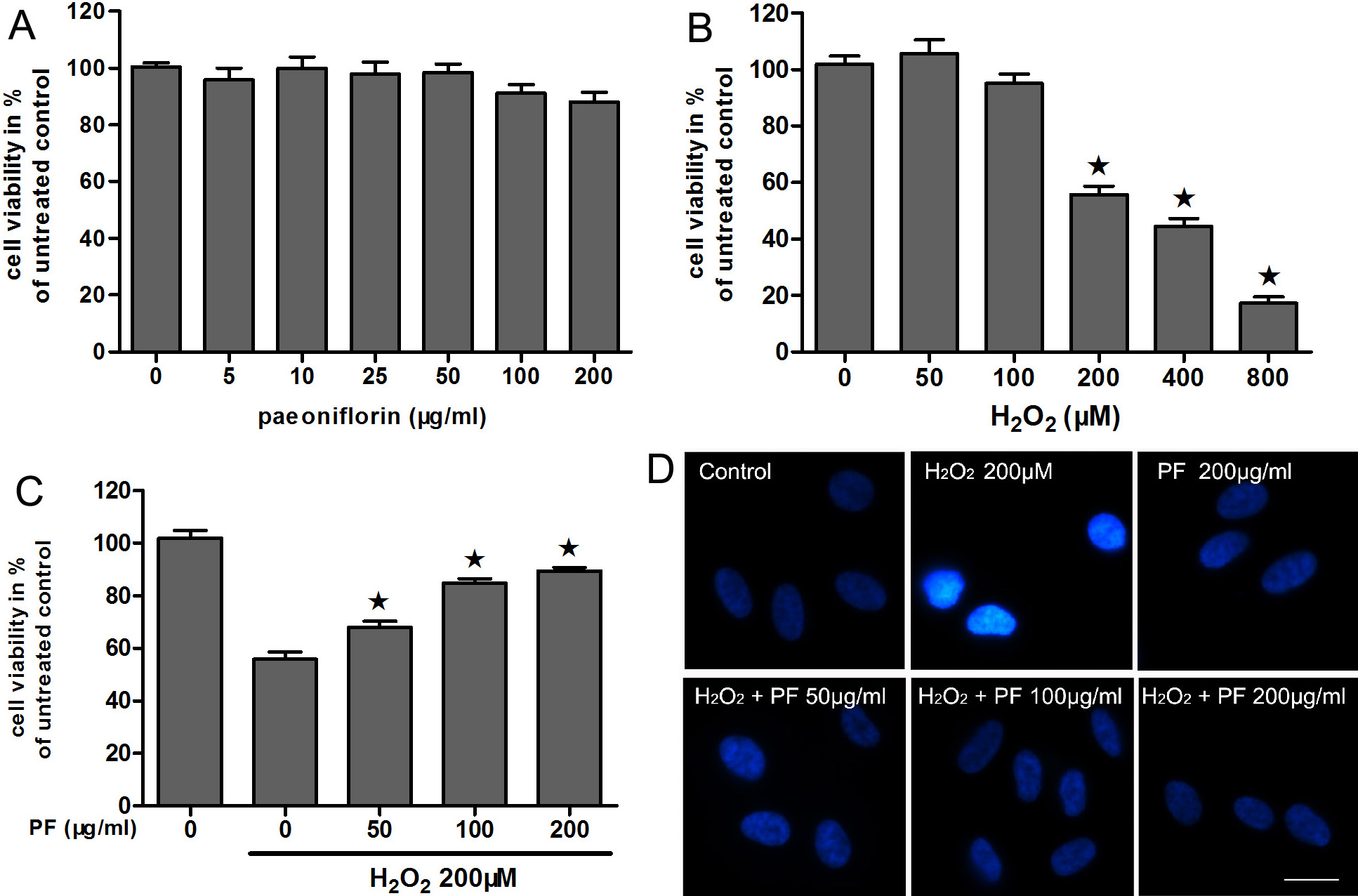
 Figure 2
of Wankun, Mol Vis 2011; 17:3512-3522.
Figure 2
of Wankun, Mol Vis 2011; 17:3512-3522.