Figure 1. Hoechst 33342 nuclei
staining and tight-junction immunostaining with zonula
occludens-1 (ZO-1) antibody illustrate the differences between
conventional immunohistochemistry on a corneal cross-section
where only a few endothelial cells are partially visible, and en
face view showing a wide field of intact cells. Original
magnification 40×. Bar 10 μm.
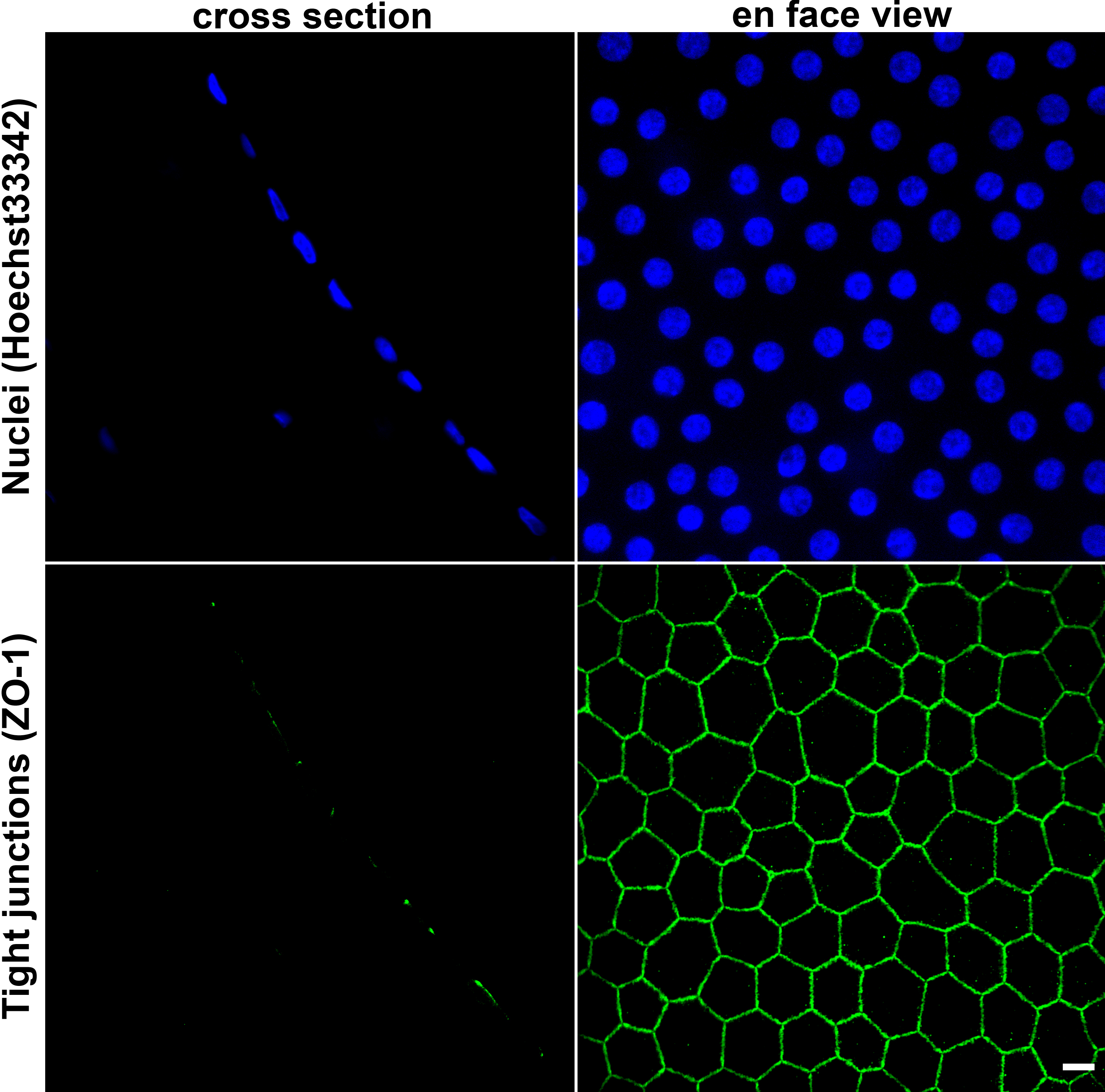
 Figure 1
of He, Mol Vis 2011; 17:3494-3511.
Figure 1
of He, Mol Vis 2011; 17:3494-3511.