Figure 1. Histopathologic features of
the eyes enucleated from experimental autoimmune uveitis (EAU),
recovery phase and control mice. A: Eye of CFA controls.
Normal retinal structure in a B10.RIII mouse. Hematoxylin-eosin
(H and E) staining (magnification, 200×). B: Eye of EAU
mice. An image from mice day 14 after IRBP immunization (at the
peak of inflammation) shows inflammatory T lymphocytes
(horizontal arrows) and macrophages (vertical thin arrow)
infiltrating the vitreous and the retina, vasculitis (vertical
bold arrow), damage to the retinal photoreceptor cell layer
(pounds) and granuloma (asterisk). H and E staining
(magnification, 200×). C: Eye of recovery phase mice. An
image from mice week 4–5 or more after IRBP immunization shows
no obvious inflammation in the retina. H and E staining
(magnification, 200×). D: Mean histopathologic score
during the development of EAU. * indicates p<0.001 when
compared EAU with control mice. Each value represents the
mean±SD (n=6).
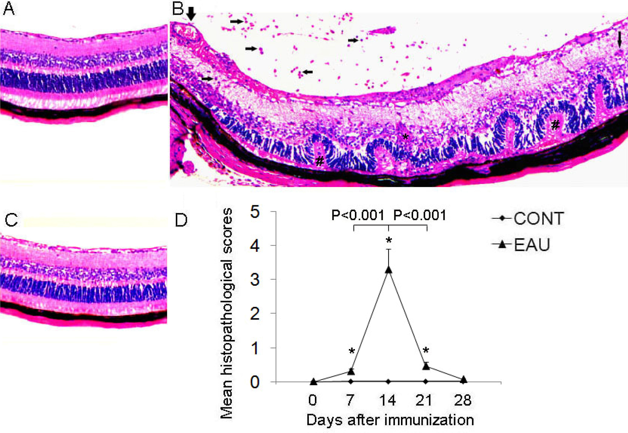
 Figure 1
of Qin, Mol Vis 2011; 17:3486-3493.
Figure 1
of Qin, Mol Vis 2011; 17:3486-3493.