Figure 5. Expression of cyclooxygenase-2 (COX-2), leukocyte common antigen (LCA), and CD3. A: COX-2-expressing cells have round and oval shapes. B: A magnified view of the outlined area of panel A. COX-2-expressing cells have oval shapes and abundant cytoplasm. Strong COX-2 staining was observed both in the cytoplasm
and in the nucleus. (arrowheads) C-D: Immunohistochemistry of adjacent sections of tissue with anti-LCA and anti-CD3 antibodies (T-cell marker) was performed.
COX-2-expressing cells (arrows 1 to 5) showed different distributions compared to inflammatory cells expressing LCA or CD3
(case 7).
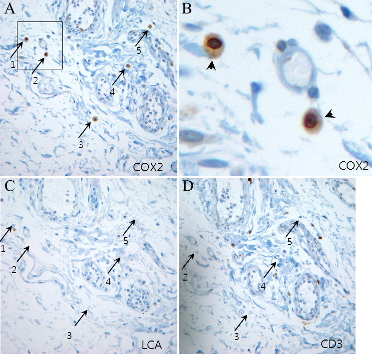
 Figure 5 of
Park, Mol Vis 2011; 17:3468-3480.
Figure 5 of
Park, Mol Vis 2011; 17:3468-3480.