Figure 1. Cyclooxygenase-2 expression
in pterygium tissue. A: Hematoxylin and eosin staining
of human pterygium (case 10) is demonstrated. B:
Immunohistochemistry of human pterygium using
anti-cyclooxygenase-2(COX-2) antibody was performed. Diffuse
expression (grade 2) of COX-2 is seen in the epithelial layer
with some strong COX-2-expressing cells in both the epithelium
and stroma. Endothelial cells lining vessels were also strongly
positive for COX-2 expression in this sample. The arrow
indicates the magnified area. C, D: COX-2
expression was not found in the epithelial or stromal layer in
the normal control. E: Strong COX-2 expression in colon
cancer was demonstrated as the positive control. F:
western blot analysis revealed various degrees of COX-2
expression in pterygium tissue (cases 2, 3, 4, 6, and 10);
however, no COX-2 band was observed in the control tissue.
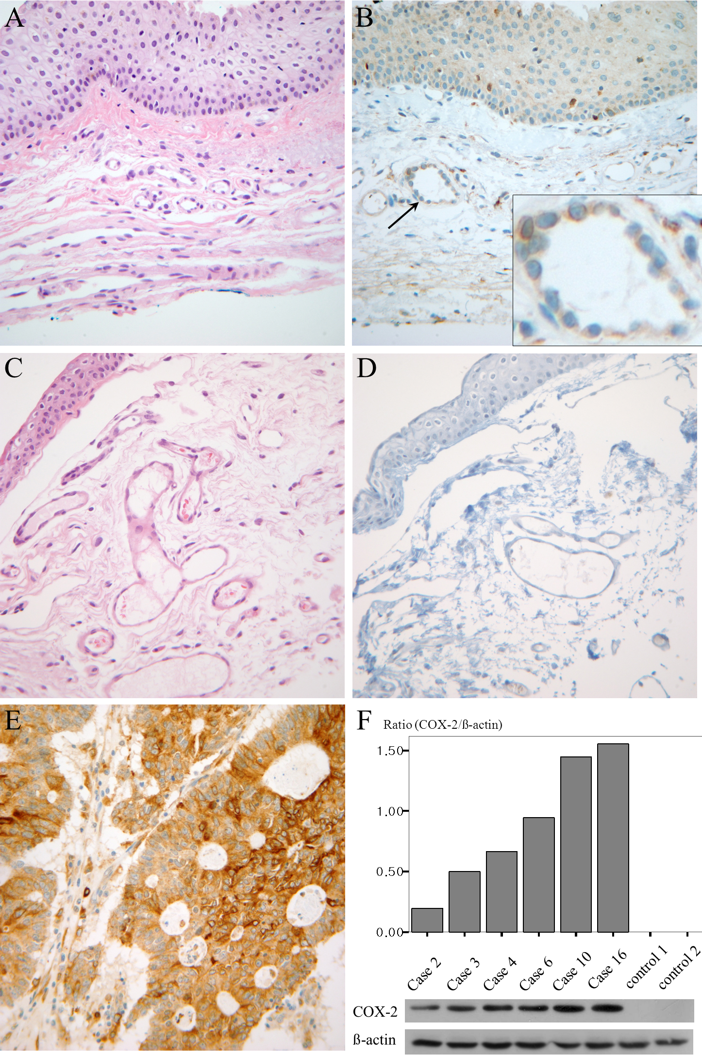
 Figure 1
of Park, Mol Vis 2011; 17:3468-3480.
Figure 1
of Park, Mol Vis 2011; 17:3468-3480.