Figure 8. Representative images of
cultured keratocytes from ReLEx (Refractive Lenticule
Extraction) lenticules. A, B, E, G:
Fresh samples. C, D, F, H:
Cryopreserved samples. A, C: ReLEx lenticule. B,
D: Free floating stromal keratocytes following enzymatic
digestion for at least 4 h in collagenase. E, F:
Attached keratocytes beginning to elongate into spindle-like
fibroblastic cells by Day 2 in culture. G, H:
Confluent stromal fibroblasts after 7 days in culture.
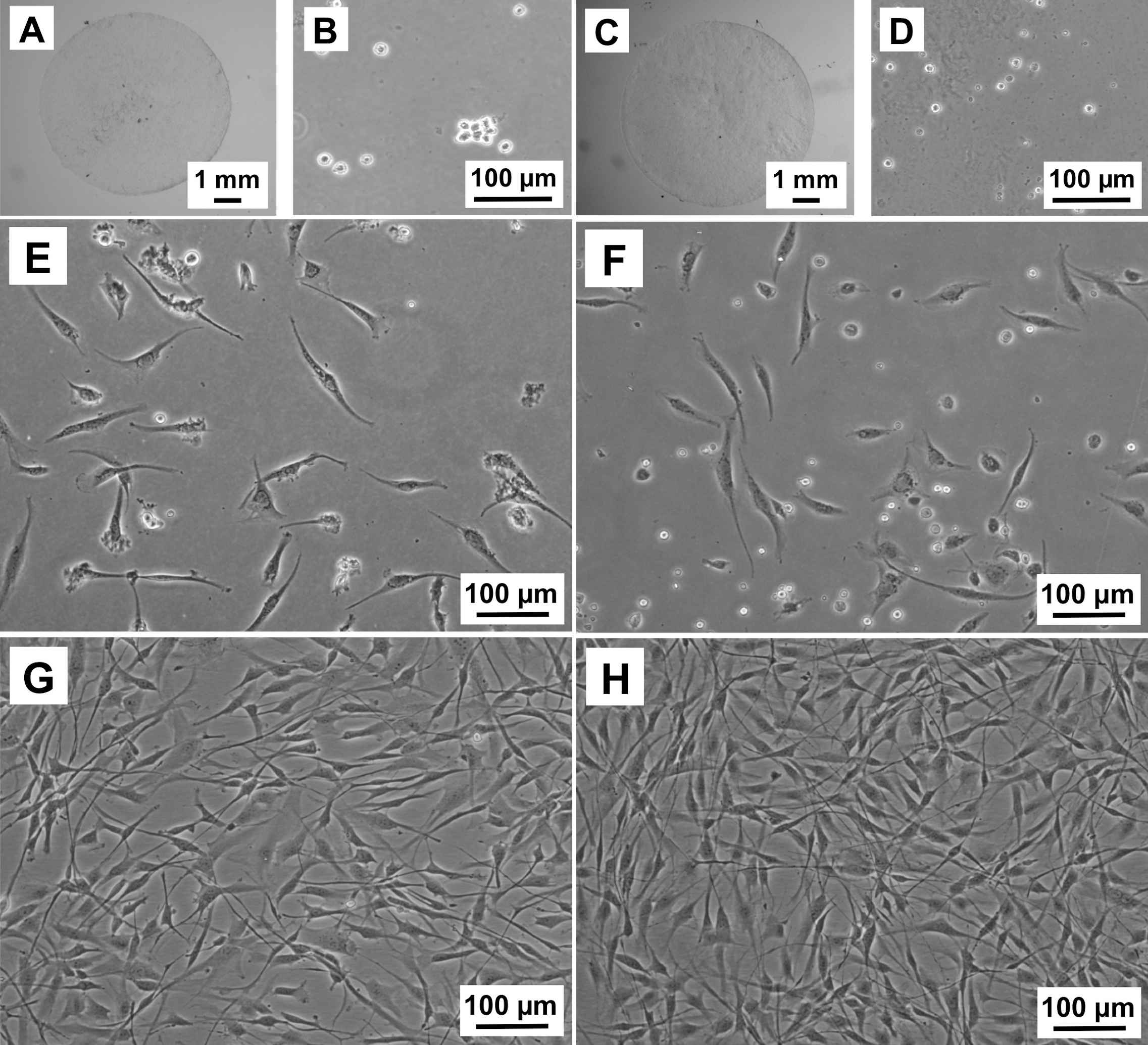
 Figure 8
of Mohamed-Noriega, Mol Vis 2011;
17:3437-3449.
Figure 8
of Mohamed-Noriega, Mol Vis 2011;
17:3437-3449.