Figure 4. Transmission electron
micrographs (TEM) of stromal lenticule showing keratocytes. A,
C: Fresh lenticule. B, D: Cryopreserved
lenticule. A, B: Apoptotic keratocytes with
cromatin condensation and fragmentation, apoptotic bodies, loss
of cytoplasm and cell shrinkage. C, D: Necrotic
keratocyte, with incomplete nuclear membrane and vacuoles in the
cytoplasm. Magnification, 8900×.
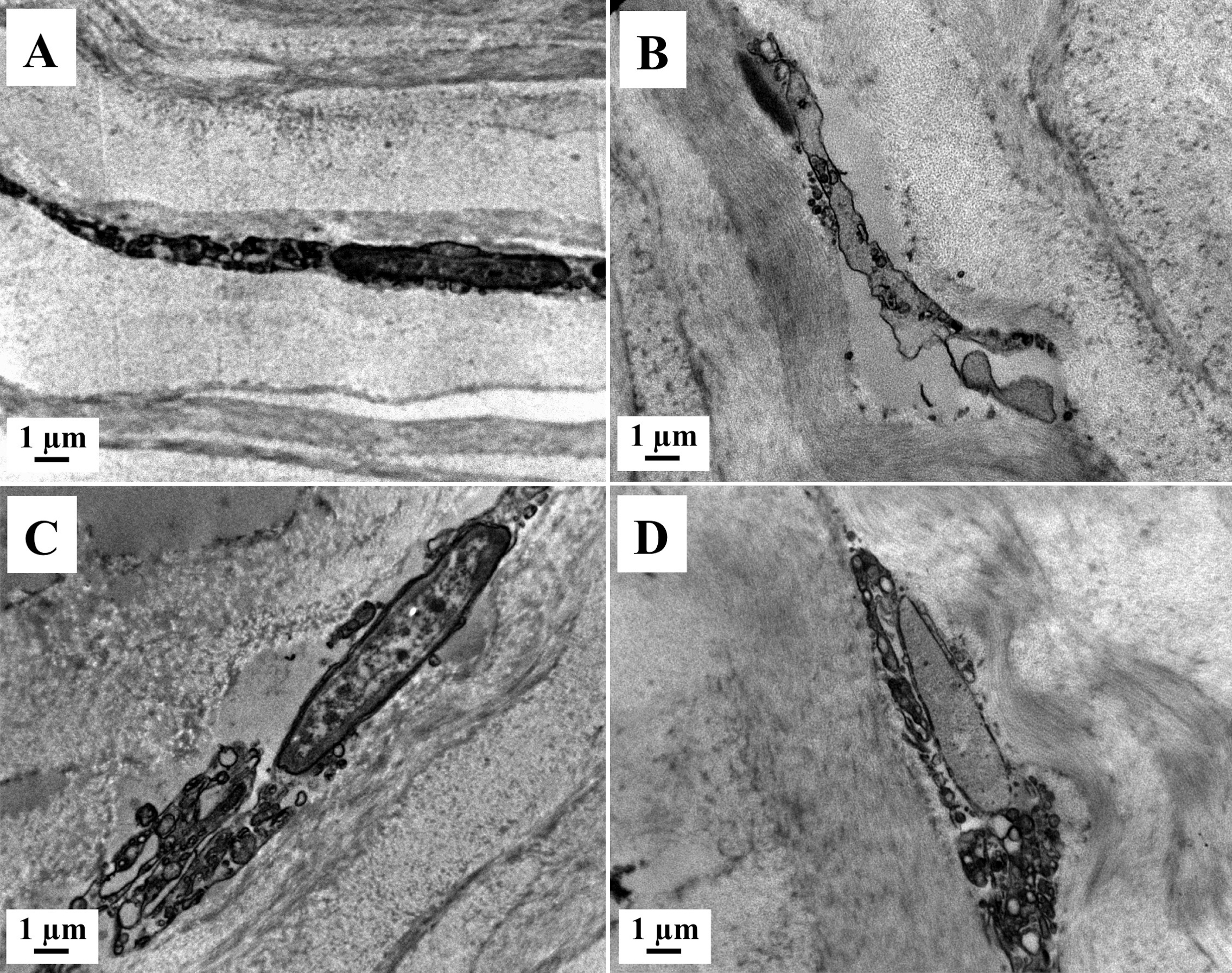
 Figure 4
of Mohamed-Noriega, Mol Vis 2011;
17:3437-3449.
Figure 4
of Mohamed-Noriega, Mol Vis 2011;
17:3437-3449.