Figure 3. Surgical steps in
Femtosecond lenticule extraction (FLEx) A: Dissection,
opening and lifting of the flap (arrow). B: Once the
flap (short arrow) is flipped, the edge of the lenticule (long
arrow) and the plane of the posterior surface of the lenticule
are identified. C: The lenticule (arrow) is separated
and removed from the cornea. D: After extraction of the
lenticule, the previous location of the lenticule edge (arrow)
can be identified in the surface of the stromal bed just before
the flap is repositioned.
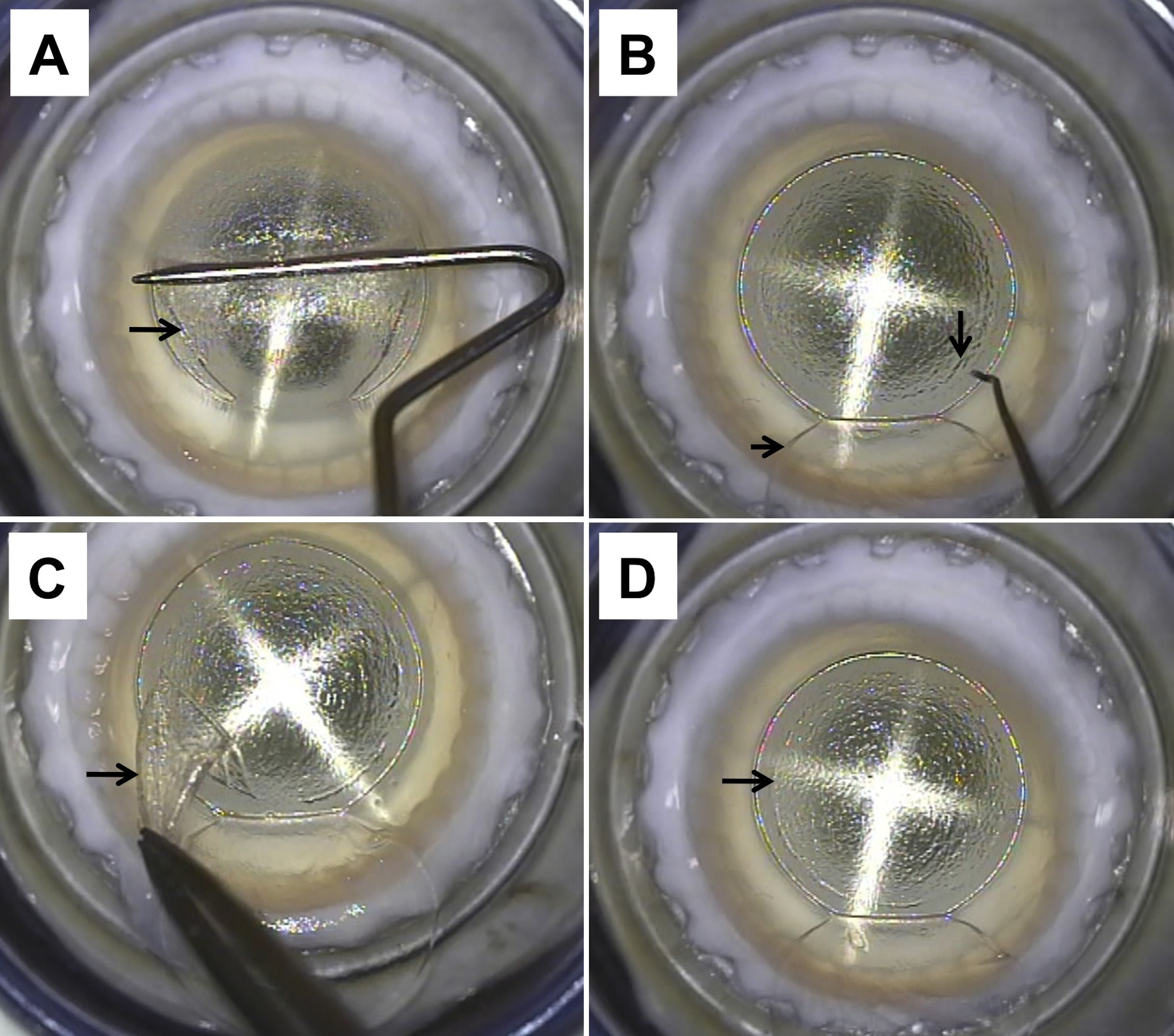
 Figure 3
of Mohamed-Noriega, Mol Vis 2011;
17:3437-3449.
Figure 3
of Mohamed-Noriega, Mol Vis 2011;
17:3437-3449.