Figure 2. Laser scanning pattern and
incisions created during femtosecond lenticule extraction (FLEx)
procedure in the human cornea. A-D:
Representative images of specific steps. A: Before the
delivery of the laser the eye must be centered and full suction
must be achieved. B: Creation of the posterior surface
of the refractive lenticule with a scanning pattern in a
centripetal direction (spiral in, arrow). C: Creation of
the anterior surface of the refractive lenticule with a scanning
pattern in a centrifugal direction (spiral out, arrow). D:
Edge of the lenticule (long arrow) and creation of the periphery
of the flap (short arrow).
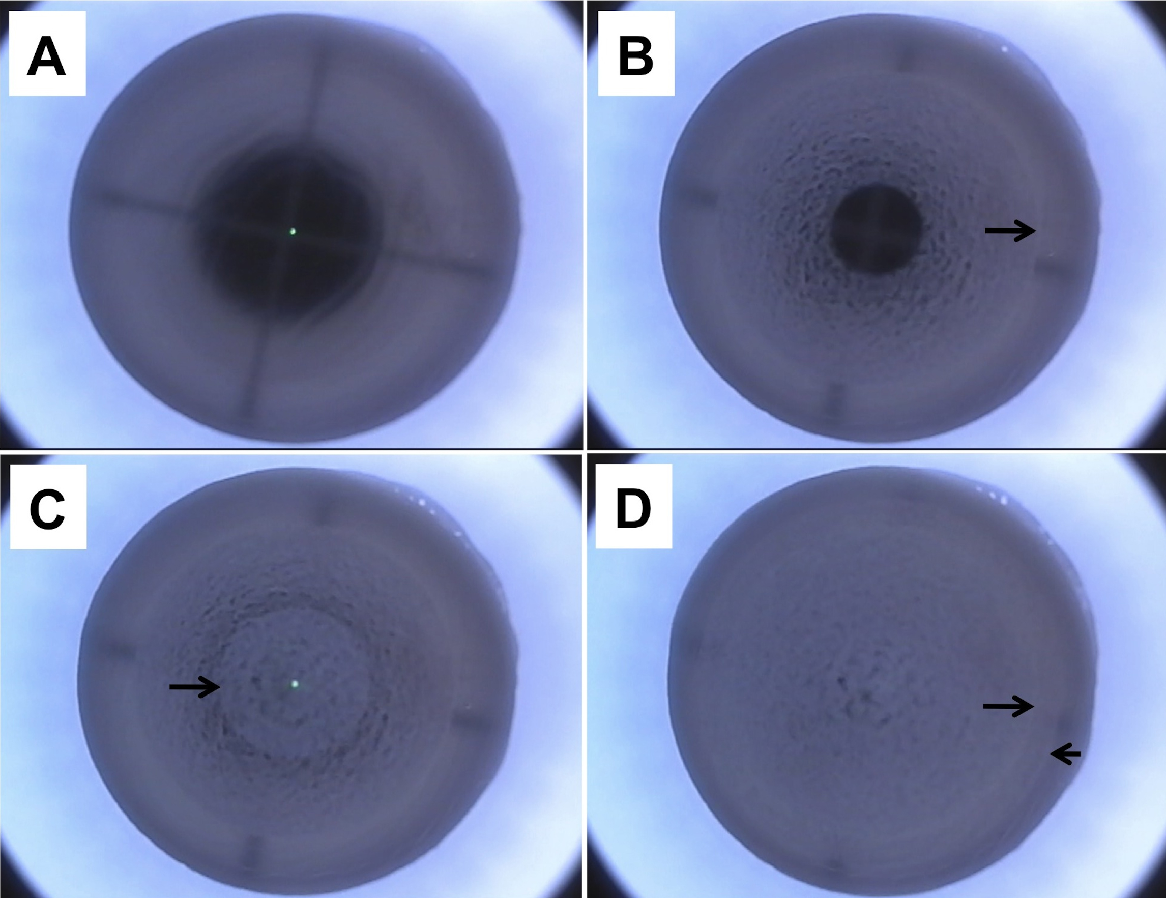
 Figure 2
of Mohamed-Noriega, Mol Vis 2011;
17:3437-3449.
Figure 2
of Mohamed-Noriega, Mol Vis 2011;
17:3437-3449.