Figure 6. Electrophysiology measurements
of three representative cases of the family studied. This figure
represents electro-oculograms (EOGs; A), right eye flash
electroretinograms (ERGs; B) and right eye multifocal
electroretinograms (mfERGs; C) in patients carrying one
mutation heterozygously (II-1: p.Y5X; III-2: p.S144G) or both mutations
(III-1). Findings are based on ISCEV standard. Patients II-2 and II-3
displayed electrophysiological findings similar to III-2 and patient
III-4 displayed electrophysiological findings similar to III-1. Except
for II-1, the amplitudes for the light phase of the EOG (A) were
abnormal with a reduction in the Arden ratio (EOG light rise <150%).
In patient III-1 (and III-4), flash ERGs show generalized decreased rod
and cone photoreceptor amplitudes and decreased photopic oscillatory
potentials amplitude (Phot-Ops; B). MfERG records a reduced
central function with relative preservation of the amplitude response
and timing from the surrounding macula (C).
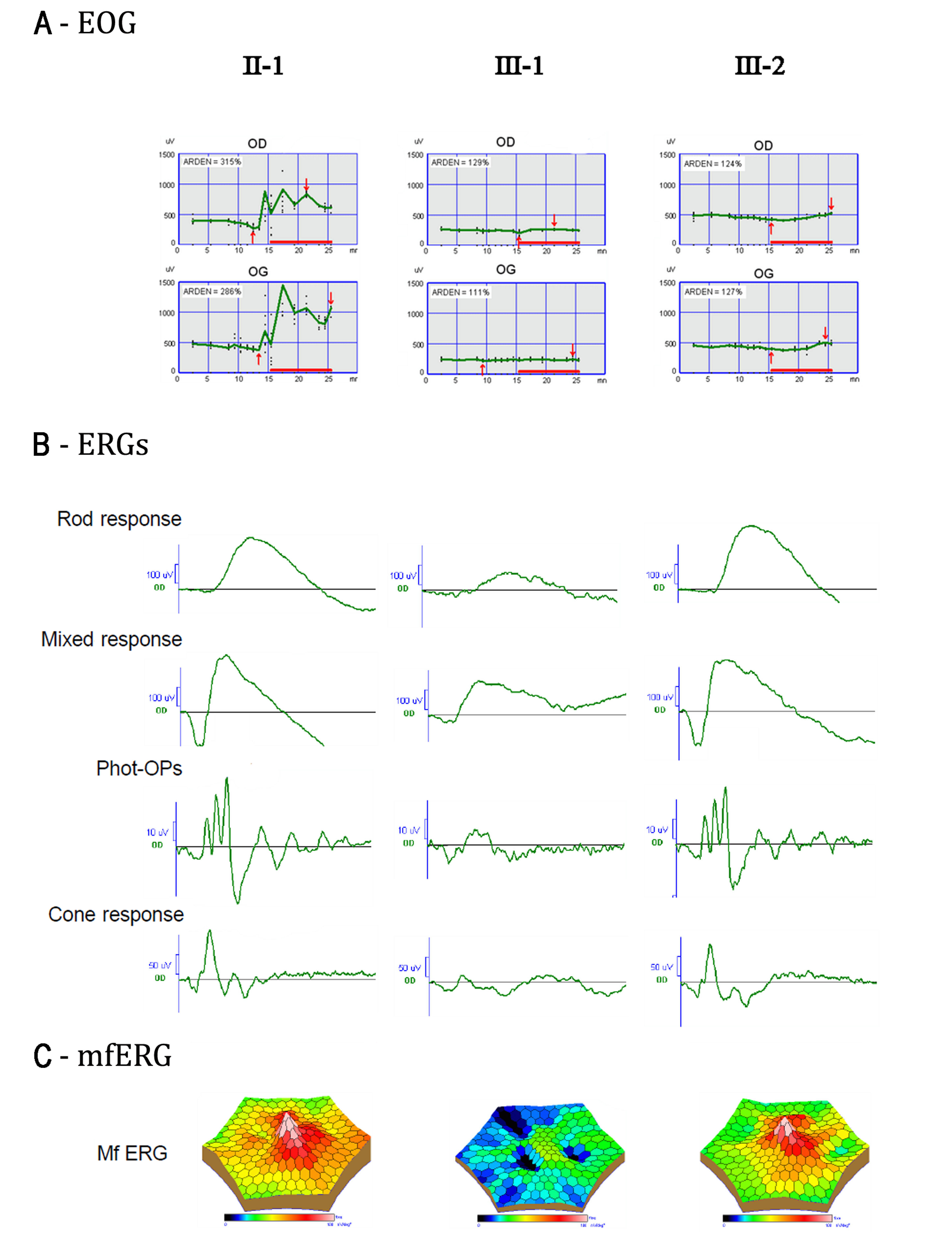
 Figure 6 of Lacassagne, Mol Vis 2011; 17:309-322.