Figure 4. Right and left eye color fundus,
autofluorescent fundus, indocyanine green angiography, and optical
coherence tomography scans in the proband (patient III-1). Well
demarcated vitelliform lesions in the central macula are detected by
fundoscopy (A-E) and are also apparent on the
autofluorescence image (B-F) and indocyanine green
angiography (C-G) in both eyes. Optical coherence
tomography images through the fovea show a highly reflective thickened
layer at the level of the retinal pigment epithelium and
choriocapillaris of both eyes and well circumscribed elevation of the
retinal pigment epithelium in both eyes (D-H).
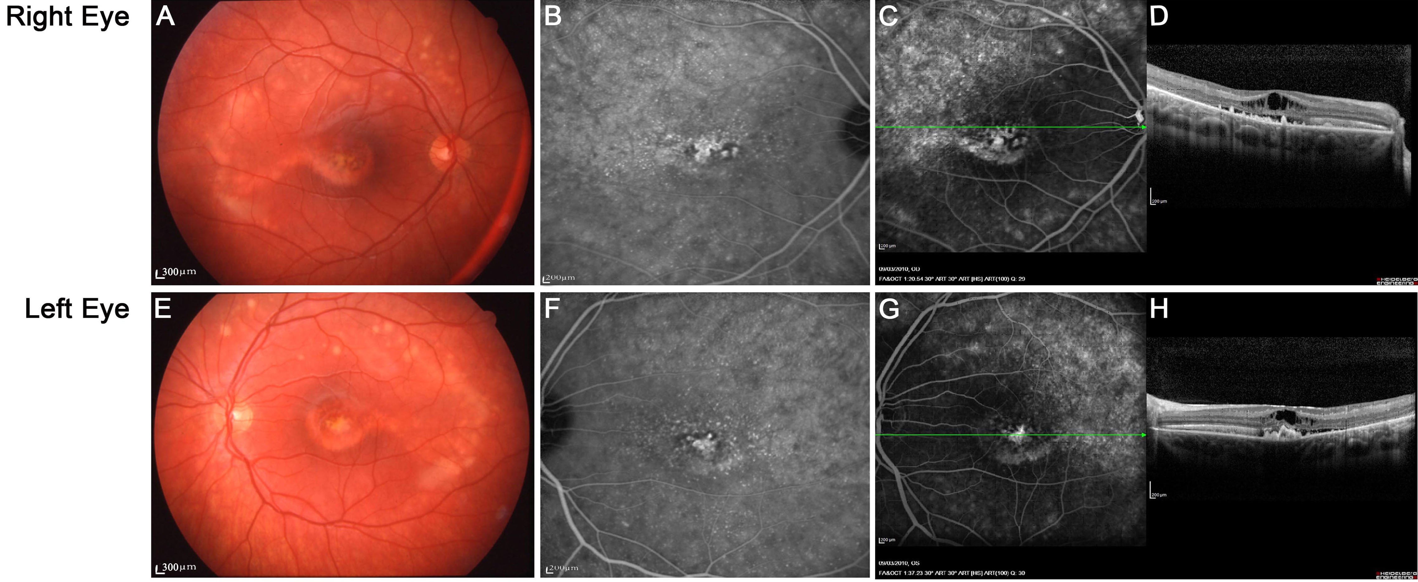
 Figure 4 of Lacassagne, Mol Vis 2011; 17:309-322.