Figure 3. Color fundus and autofluorescent
fundus/optical coherence tomography scans in patients II-1, II-2, II-3,
III-2, and III-3. No macular lesion was detectable by autofluorescence
imaging or optical coherence tomography scanning for patients II-1 or
III-2. No macular lesion was detectable in the color fundus for
patients II-2, II-3, or III-3.
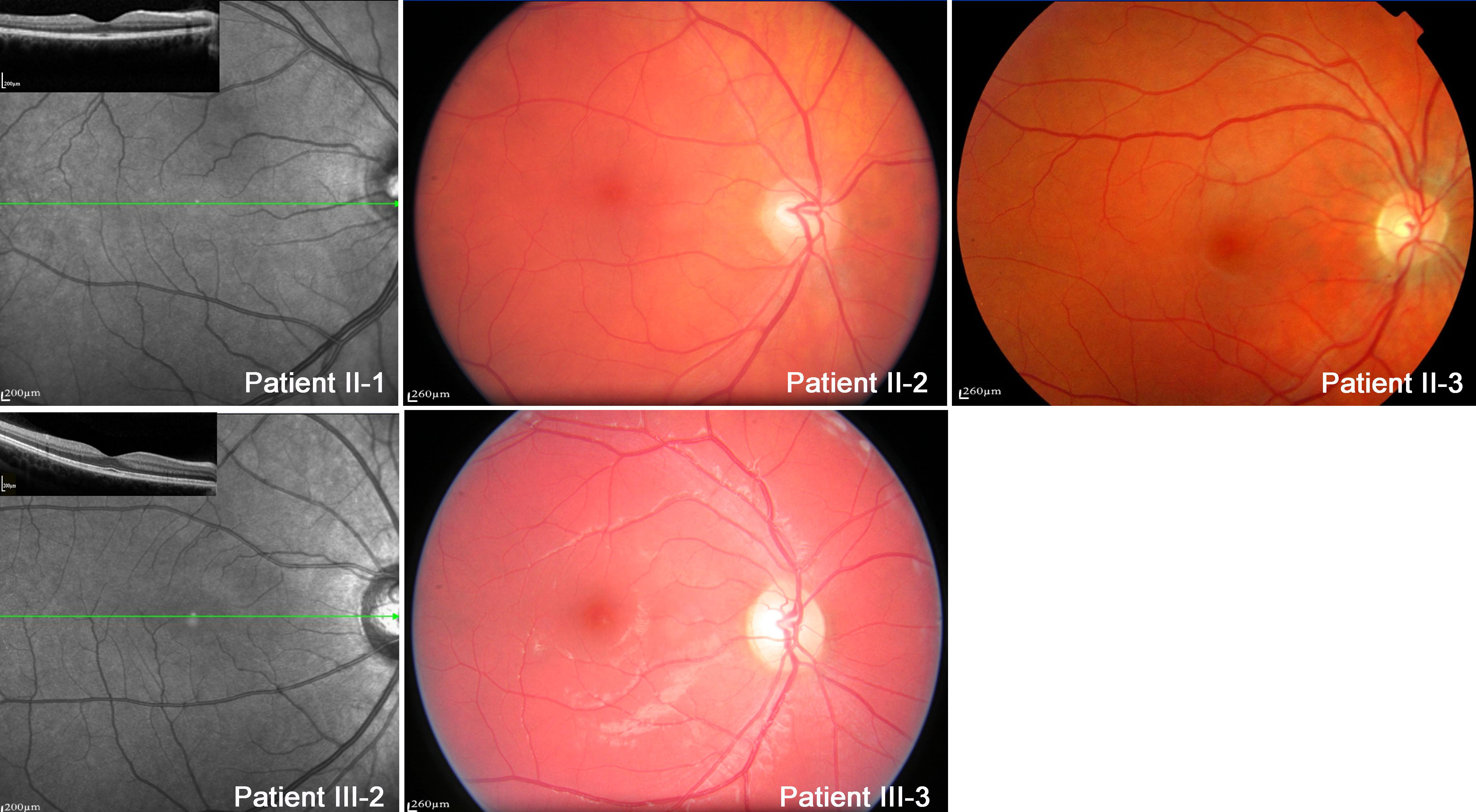
 Figure 3 of Lacassagne, Mol Vis 2011; 17:309-322.