Figure 8. Native page of crystallins. A: Native page showed the bands of native α-crystallin isolated from lenses exposed to 0–100 µM Dex located on the top of the
page and HMW of α-crystallin were larger than 700 kDa. Native β-crystallin located on the middle of the page. B: Expressional levels of α-crystallin increased after lens was exposed to 1 µM Dex, β-crystallin increased after lens was
exposed to Dex (*<0.05 means significantly). C: Analysis of percentages of native crystallins showed percentage of α-crystallin gradually decreased and β-crystallin gradually
increased with increasing concentrations of Dex.
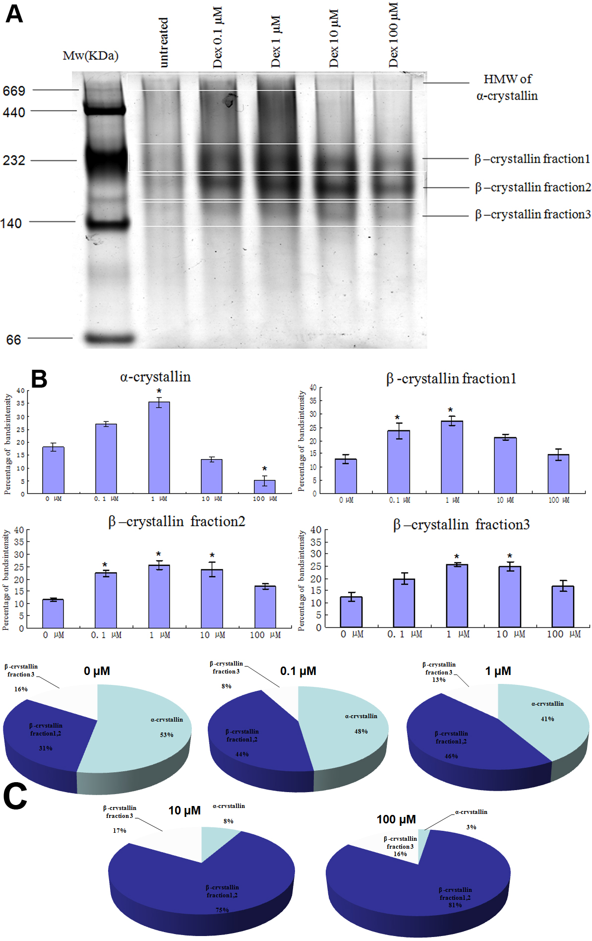
 Figure 8 of
Wang, Mol Vis 2011; 17:3423-3436.
Figure 8 of
Wang, Mol Vis 2011; 17:3423-3436.