Figure 7. EM showed some changes in lens without or with Dex exposed lenses. A: Clear lens exposed to 0 µM Dex. B: Circular lesions were found located between the lens fiber cells. C: Showing increased intercellular space, circular lesions, spherical hyperplasia denaturation were found in mitochondrial
of fiber cells. D: Showing irregular arrangement of lens fiber cells, cristae swelling and medullary sheath change in mitochondrial. E: Seriously increased intercellular space was found after the lens was exposed to 100 µM Dex.
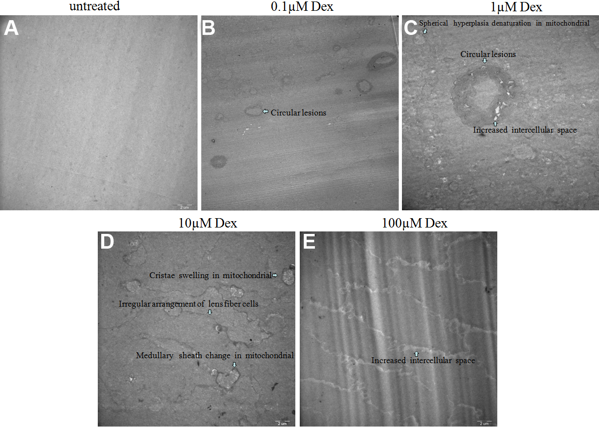
 Figure 7 of
Wang, Mol Vis 2011; 17:3423-3436.
Figure 7 of
Wang, Mol Vis 2011; 17:3423-3436.