Figure 6. Histology of lenses without or with dexamethasone-exposed lenses. Whole lenses were cultured for 48 h without (control) or
with dexamethasone. Lens sections were stained with primary antibodies(same with western-blot experiment) then stained with
a second round of antibodies (HRP-conjuncted,Cy3 or FITC-conjuncted).
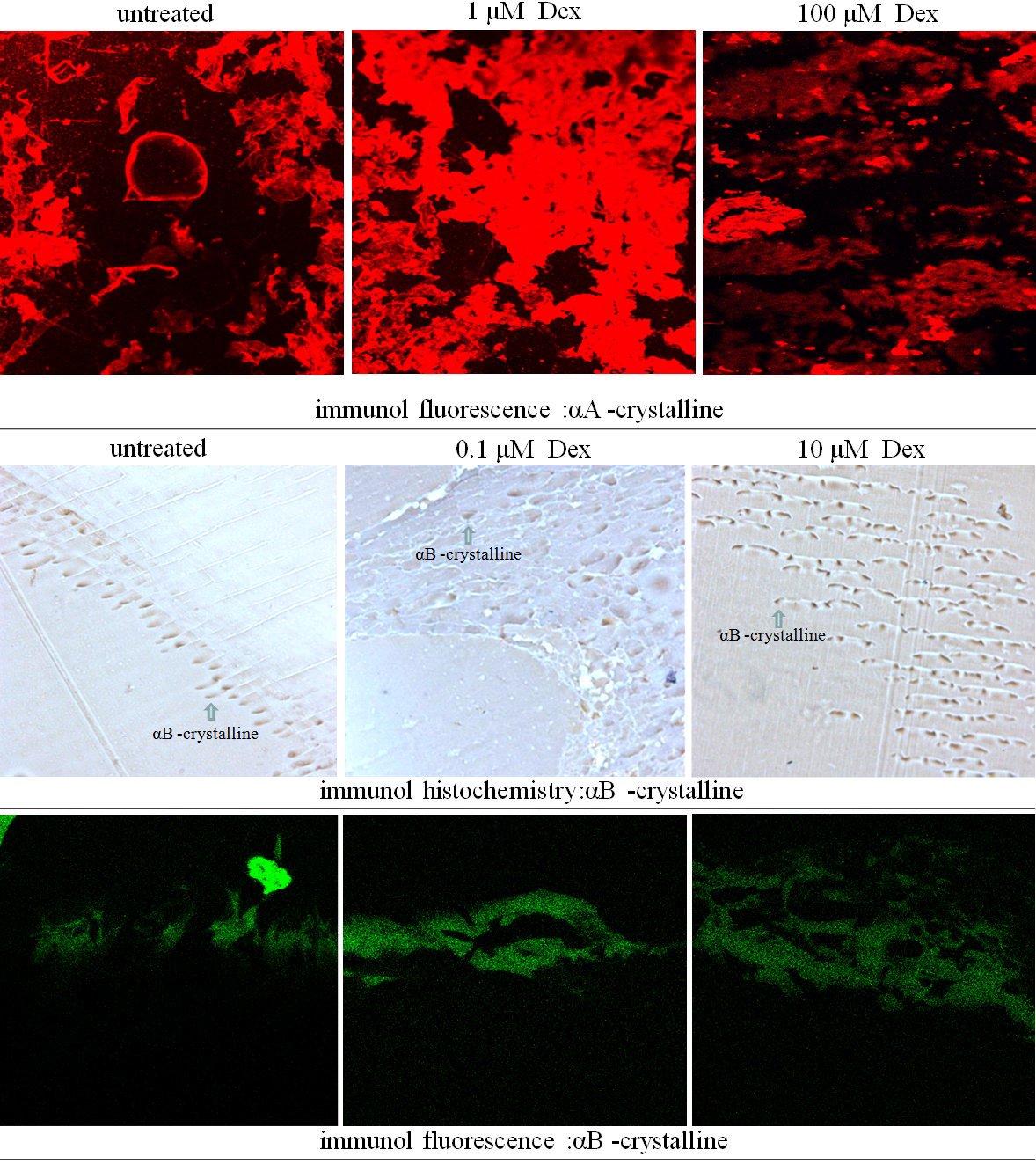
 Figure 6 of
Wang, Mol Vis 2011; 17:3423-3436.
Figure 6 of
Wang, Mol Vis 2011; 17:3423-3436.