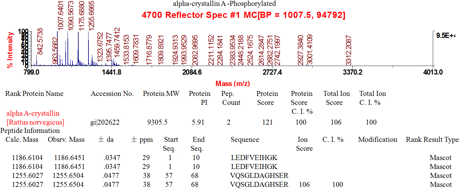
Figure 4. Identification of rat
phosphorylated-αA-crystallin, by mass spectrometry of the
protein spot digested with trypsin. Amino acid sequences of
tryptic peptides recovered from the protein spots were presented
in two-dimensional gel electrophoretic maps of water-
insoluble-urea-soluble protein fraction, isolated from the lens
after exposure to variable concentrations of Dex. The amino acid
sequences reported are the tryptic fragments from individual
spot, and they did not alter compared with amino sequences of
proteins published in
PubMed.
Thus, we present the MS spectra (PMF spectra) of it.
 Figure 4
of Wang, Mol Vis 2011; 17:3423-3436.
  Figure 4
of Wang, Mol Vis 2011; 17:3423-3436.