Figure 9. Effects of progesterone on
IL-1β–induced MAPK and IκB-α phosphorylation in corneal
fibroblasts. Cells were incubated in the absence or presence of
progesterone (10 μM) for 12 h and then in the additional absence
or presence of IL-1β (0.1 ng/ml) for 30 min. Cell lysates were
then prepared and subjected to immunoblot analysis with
antibodies to total or phosphorylated (P-) forms of ERK, JNK,
p38 MAPK, or IκB-α. Data are representative of three independent
experiments.
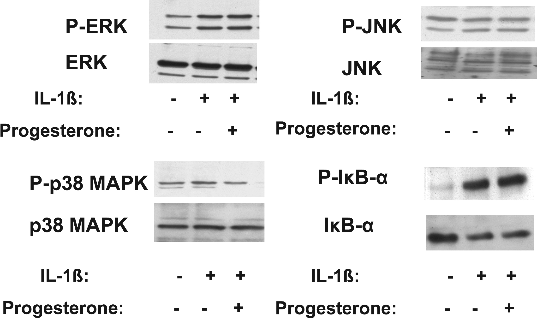
 Figure 9
of Zhou, Mol Vis 2011; 17:3415-3422.
Figure 9
of Zhou, Mol Vis 2011; 17:3415-3422.