Figure 2. Immunofluorescence images.
A: In the no-scratch group, after cell confluency,
vascular endothelial (VE)-cadherin yielded continuous, linear
staining around the periphery of EA.hy926 cells (confocal
microscopy 200×). B: In the no-scratch group, after cell
confluency, there was no expression of vimentin protein
(confocal microscopy 100×). C: In the no-scratch group,
after cell confluency, there was no expression of the Twist
protein (confocal microscopy 100×). D: In the scratch
group, the VE-cadherin expression of the endothelial cells that
migrated into the scratch wound was decreased at endothelial
cell-cell junctions and was mainly localized to the cytoplasm;
VE-cadherin yielded continuous, linear staining around the
periphery of EA.hy926 cells on the outside of the scratch wound
(confocal microscopy 100×). E: In the scratch group, the
vimentin expression of endothelial cells that migrated into the
scratch wound (confocal microscopy 100×). F: In the
scratch group, the Twist expression of endothelial cells that
migrated into the scratch wound (confocal microscopy 100×).
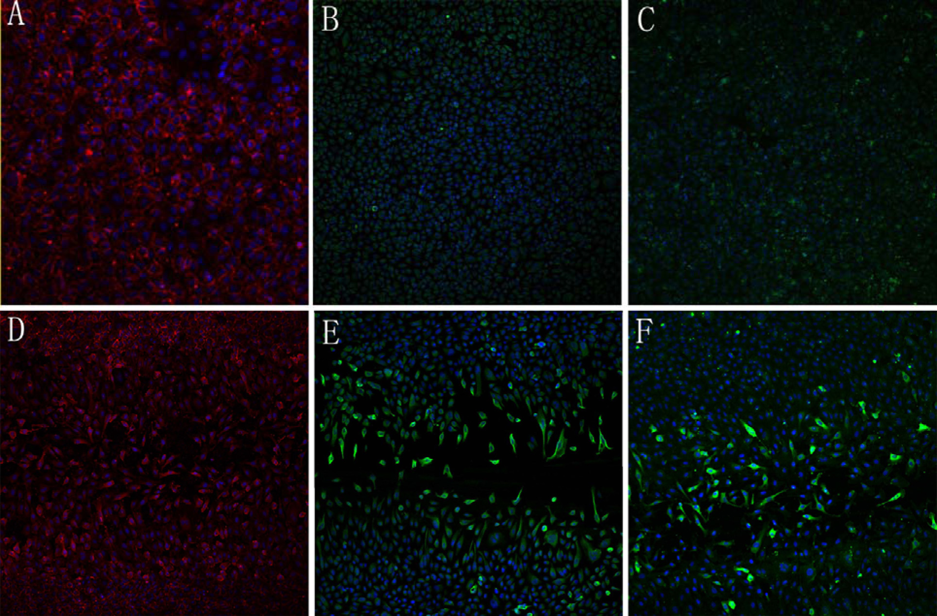
 Figure 2
of Gao, Mol Vis 2011; 17:3406-3414.
Figure 2
of Gao, Mol Vis 2011; 17:3406-3414.