Figure 2. Immunofluorescent staining detect the protein expression of TLR2 and TLR4. The high glucose and high mannitol stimulated cells
treated with anti-TLR2 (A) and anti-TLR4 (B) antibodies and stained by Cy3 and DAPI dihydrochloride. There was no immunoreactivity in the negative control (isotype IgG).
Merge means overlap the DAPI and Cy3.
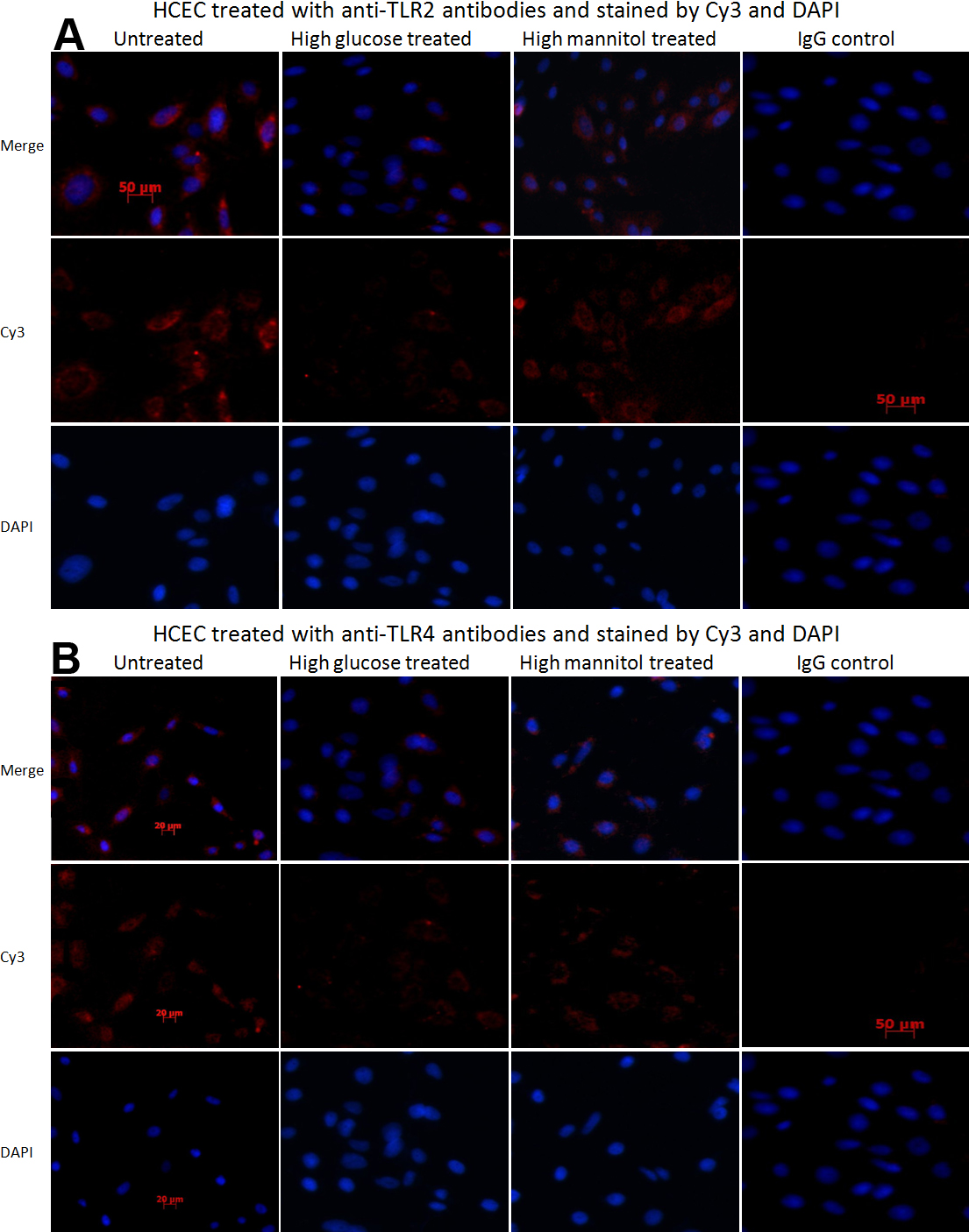
 Figure 2 of
Ni, Mol Vis 2011; 17:3384-3391.
Figure 2 of
Ni, Mol Vis 2011; 17:3384-3391.