Figure 6. An anterior-posterior
maturation gradient is maintained through development of the
iris from the OCL. A, C-I: Transverse
sections through E14 eyes. A: A section labeled with
Pax3 and MF20. MF20 is absent from the mesenchyme closest to the
OCL (bracket). Pax3/MF20 are expressed in the same cells at the
anterior position in the skeletal muscle (arrows). B: A
confocal image of a wholemount E16 eye labeled with Pax6 and
MF20. Bracket indicates a Pax only zone in the eye anterior to
musculature. C: A section labeled with MF20. The bar
indicates the iris sphincter muscle and the anterior most 100
µm. D-I: Sections through the eyes of embryos
exposed to BrdU for 4 h (D-F) or 8 h (G-I)
before harvesting. Skeletal muscle is labeled with MF20 (D,
F, G, I) and arrows indicate myonuclei
that have incorporated BrdU (E, F, H, I).
J: Graphic summary of BrdU pulse data. There is a
significant difference in the percent of nuclei that have
incorporated BrdU comparing the anterior and posterior muscle
after an 8 h (p<0.01) but not 4 h (p>0.05) pulse. Scale
Bars: 100 µm, * indicates anterior of cup/anterior of pupillary
margin). L; lens, OCL; Optic Cup Lip.
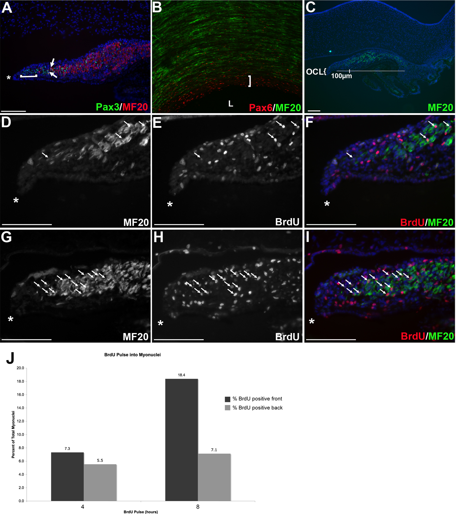
 Figure 6
of Venters, Mol Vis 2011; 17:3347-3363.
Figure 6
of Venters, Mol Vis 2011; 17:3347-3363.