Figure 5. The OCL contributes cells
throughout the extent of the eye. A-C: Wholemount
view of an E17 embryo where the OCL was infected focally with
GFP expressing replication-incompetent virus at E3. A:
The embryo head before dissection to expose the eye. B:
GFP expression in the eye of the embryo after removal of the
eyelids and cornea. GFP can be seen from the OCL at the lens (L)
to the cut edge of the eyelid (asterisk). C: Higher
magnification view of B, showing retention of GFP
expressing cells at the OCL. Bright cells have delaminated from
the OCL into the overlying mesenchyme (bracket). D: Low
magnification view of a section through the GFP expressing
region of ciliary body and neural retina of the embryo shown in
A-C. GFP is present in the ciliary folds and in
the neural retina. The posterior extent of GFP expression is
indicated (arrow). GFP is present in the RPE at a posterior
position (double arrowhead). E: Higher magnification of
the boxed area in D. GFP expressing cells in the RPE are
indicated (double arrow). F-R: Adjacent sections
through the embryo in A-C. F-H:
GFP expressing cells are co-labeled with Visinin in the neural
retina (F). G-H: GFP alone (G) and
combined with the bright-field image (H). GFP is present
in the inner neural retina layer and the overlying pigmented
epithelium (arrow). I-J: GFP and Visinin at the
CMZ. GFP is present in the pars plana of the ciliary body, the
CMZ (arrow), and in photoreceptors at the peripheral neural
retina (arrowhead). K-L: GFP is co-localized with
Collagen IX expressing tissue in the inner layer of the ciliary
folds and is also present in the outer pigmented ciliary
epithelium (arrowhead; L). M: GFP is present in
the smooth-muscle in the mesenchyme of the iris (arrowheads). N:
GFP is present in the iris mesenchyme in an MF20 expressing
myocyte (arrow) O: GFP is colocalized with Pax3
expressing cells in the iris mesenchyme (arrows). P-R:
GFP and Pax6 colocalize (arrow) in cells delaminating from the
OCL into the mesenchyme overlying the iris epithelium and in the
epithelial layers of the iris (white arrowhead) and ciliary
folds (red arrows). Q: A higher magnification of P.
R: Corresponding bright-field image with GFP expression.
Scale Bars: 100 µm. The anterior eye is to the left in all
sections except B and D. *; OCL, L; lens, CB;
ciliary body, CMZ; ciliary margin zone, NR; neural retina, PE;
pigmented epithelium, ONL; outer nuclear layer, INL; inner
nuclear layer, IPL; inner plexiform layer, GCL; ganglion cell
layer.
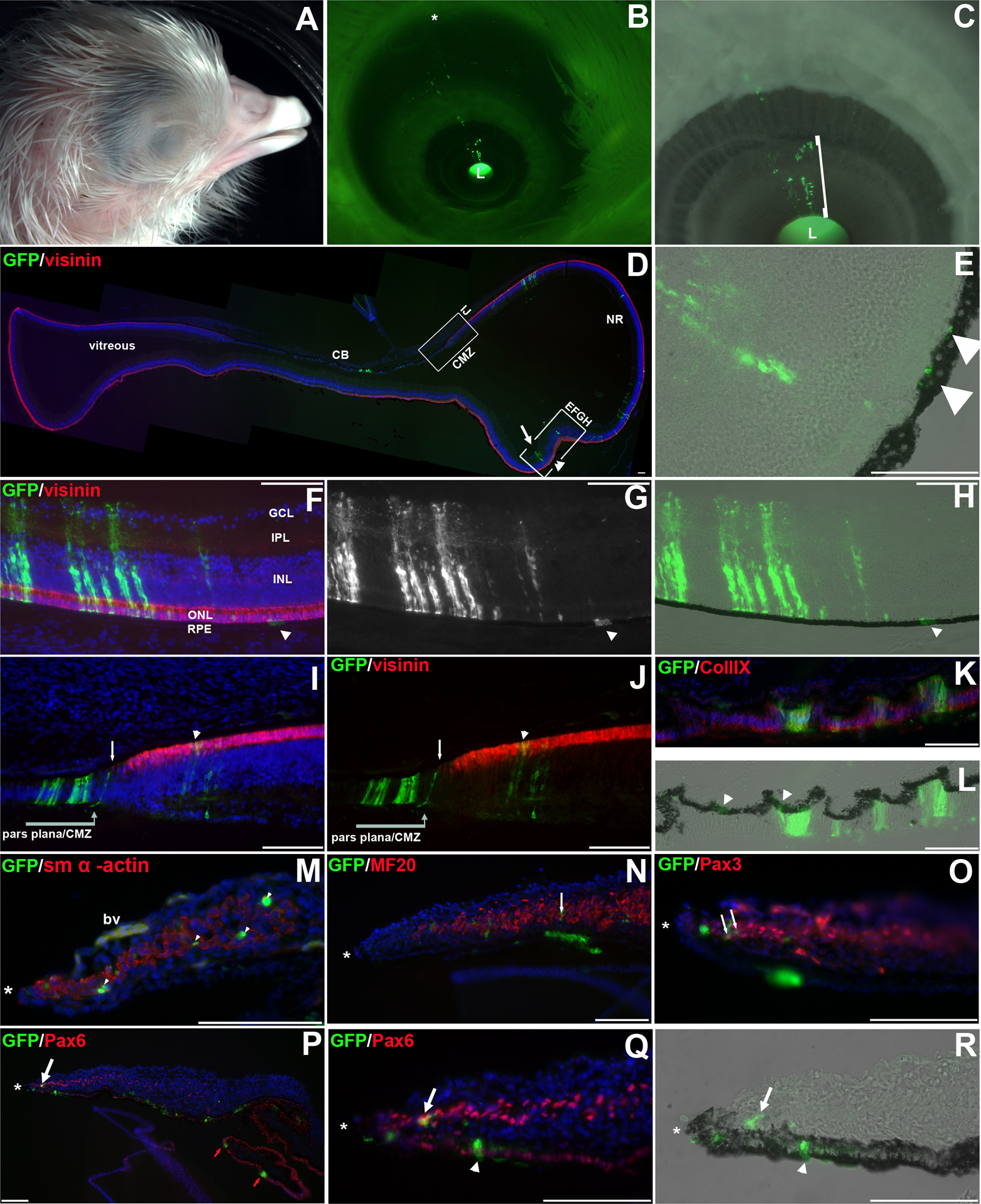
 Figure 5
of Venters, Mol Vis 2011; 17:3347-3363.
Figure 5
of Venters, Mol Vis 2011; 17:3347-3363.