Figure 4. The early optic cup lip
expresses mature fate markers. A-F: Transverse
sections through eyes of 31 somite (A-C) and 38
somite (D-G) embryos labeled with Collagen IX (A,
B, D) or NaK-ATPase (C, E, F).
A: A low magnification of the section shown in B.
B, D: Collagen IX (red) is present in the inner
layer of the anterior optic cup. The arrows mark the posterior
limit of Collagen IX expression. C, E, F:
NaK-ATPase is present in the pigmented epithelium and the OCL. F:
Expression of NaK-ATPase extends into the anterior of the
non-pigmented, inner layer in a 38 somite embryo (arrow). G:
Sox2 expression is present in the inner layer of the optic cup.
Arrow marks the anterior limit of Sox2 expression. H:
Transverse section through the ventral optic cup of a HH23
embryo. Hu is expressed at the posterior neural retina
(bracket). The posterior limit of Collagen expression in the
anterior eye is indicated (arrow). Scale Bars: 100 µm. HH;
Hamburger Hamilton Stage, Coll IX; Collagen IX, NaK; NaK-ATPase.
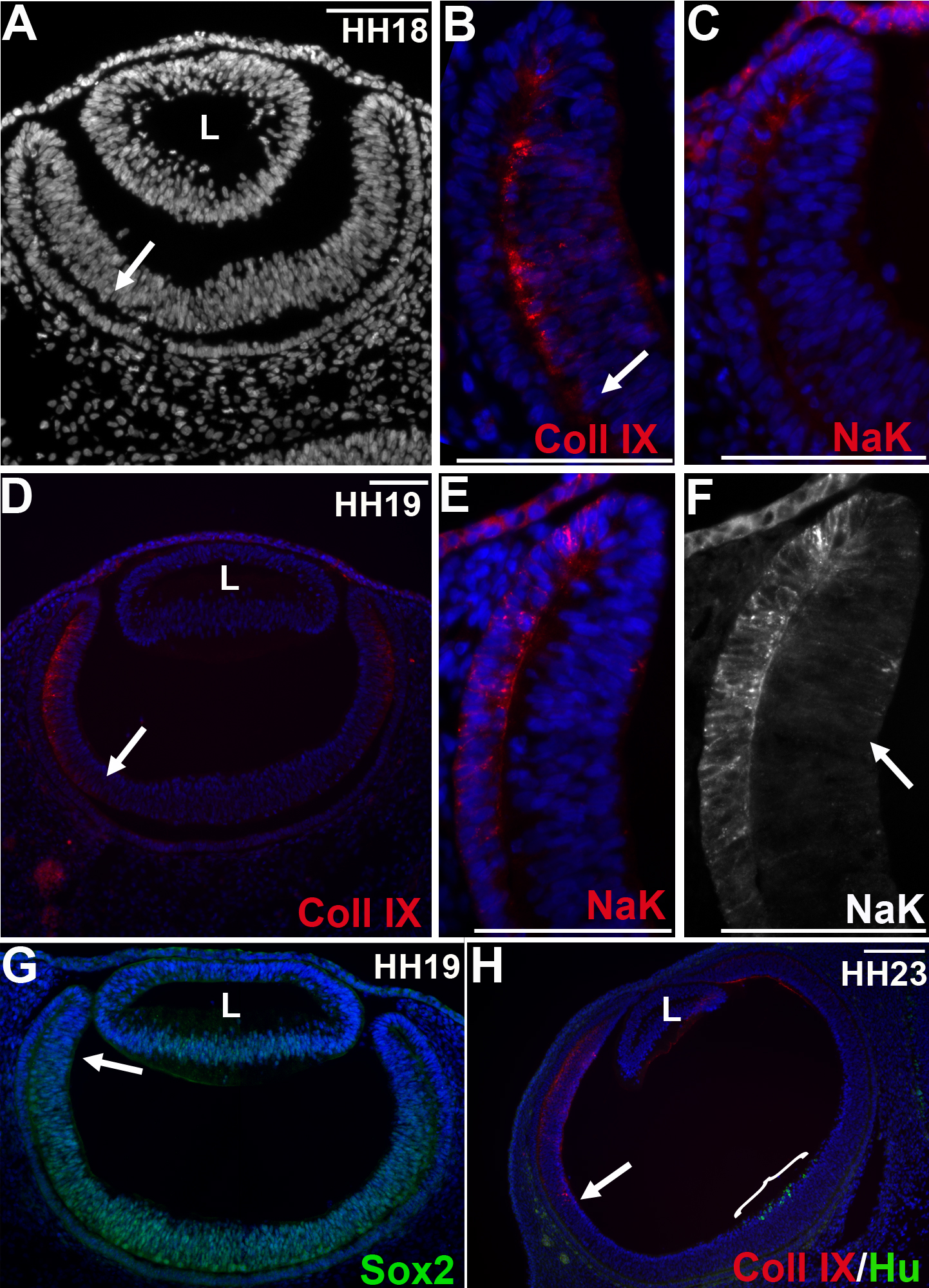
 Figure 4
of Venters, Mol Vis 2011; 17:3347-3363.
Figure 4
of Venters, Mol Vis 2011; 17:3347-3363.