Figure 2. Cells derived from the OCL
are distributed through the peripheral neural retina and ciliary
body. A-C: Whole mount views of eyes infected
with GFP-expressing replication-incompetent retrovirus targeted
to the OCL. Embryos were reincubated until E4.5 (A), E6.5
(B), and E9 (C). A: GFP expressing cells
are evident as a spoke-line emanating from the OCL adjacent to
the lens. B, C: The GFP expressing cells
traverse the morphological boundary between the ciliary body and
the peripheral neural retina (arrows, B and C). D-K:
Coronal sections through the GFP expressing region of an OCL
targeted eye. D: Phase contrast image of the eye with
relevant features indicated. Boxed area represented at higher
magnification in panels E-K. E: GFP is
evident at the OCL (arrow), in the Collagen IX expressing
ciliary body (red signal), posterior to the Collagen IX zone in
the optic epithelia and axons (red arrows). Double arrowheads
indicate the border between ciliary body and neural retina. F:
GFP expressing cells are located in the hinge at the OCL (arrow)
and in the anterior Collagen IX expressing region. G:
GFP expressing cells are seen throughout the presumptive ciliary
body. H: GFP expressing cells are located at the
posterior ciliary body/ peripheral NR, where Hu expression is
evident in a few cells (arrowheads). Blood vessels (bv) are
autofluorescent. I: GFP expressing cells are present in
the Hu expressing neural retina and GFP labeled axons are
apparent (red arrows). J: GFP is also present in the
outer, pigmented epithelium (arrows) over the neural retina. K:
Progeny of cells (different embryo) infected with a virus
encoding nuclear localized GFP show abundant GFP expression in
the pigmented epithelium overlying the neural retina (arrows).
Scale Bars: 100 µm. The anterior of the eye is to the left. OCL;
Optic Cup Lip, L; lens, NR; neural retina, CB; ciliary body, bv;
blood vessels.
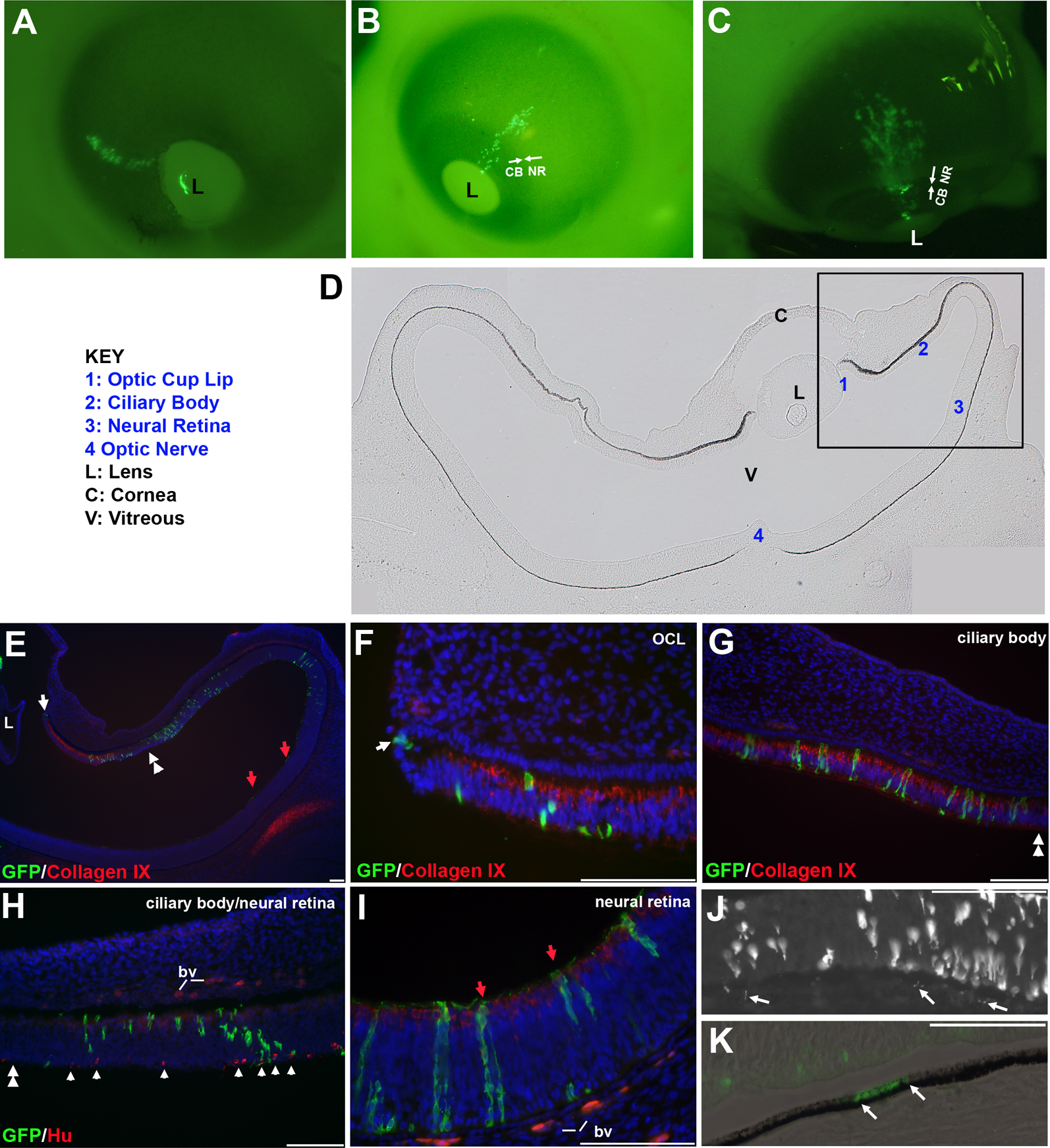
 Figure 2
of Venters, Mol Vis 2011; 17:3347-3363.
Figure 2
of Venters, Mol Vis 2011; 17:3347-3363.