Figure 3. Effect of Bevacizumab on BCE cell death as quantified by flow cytometry. A: Flow cytometry analysis of BCE cells treated with Bevacizumab and/or VEGF. Cells were stained with propidium iodide (PI)
before flow cytometry analysis. B: Viable percentage of gated cells (Non-PI staining) from experiment shown in A. BEV (Bevacizumab); V (vascular endothelial growth factor; VEGF).
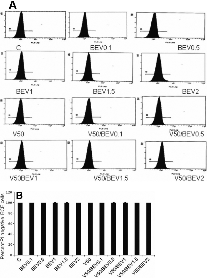
 Figure 3 of
Rusovici, Mol Vis 2011; 17:3339-3346.
Figure 3 of
Rusovici, Mol Vis 2011; 17:3339-3346.