Figure 4. Fundus photos of the four members of the family. The mother (I:2), with digenic mutations, had a normal fundus appearance.
All three patients (II:1, II:2, and II:3) from the family had similar fundus changes, including waxy, pale optic discs, artery
attenuation, generalized carpetlike retinal degeneration, macular atrophy, nummular pigmentation at the posterior pole, and
irregular pigmentation and white dots in the midperipheral region. The fundus changes in the proband (II:1) with triallelic
mutations in CRB1 and SPATA7 were similar to the those of the other two patients (II:2 and II:3) with compound heterozygous CRB1 mutations.
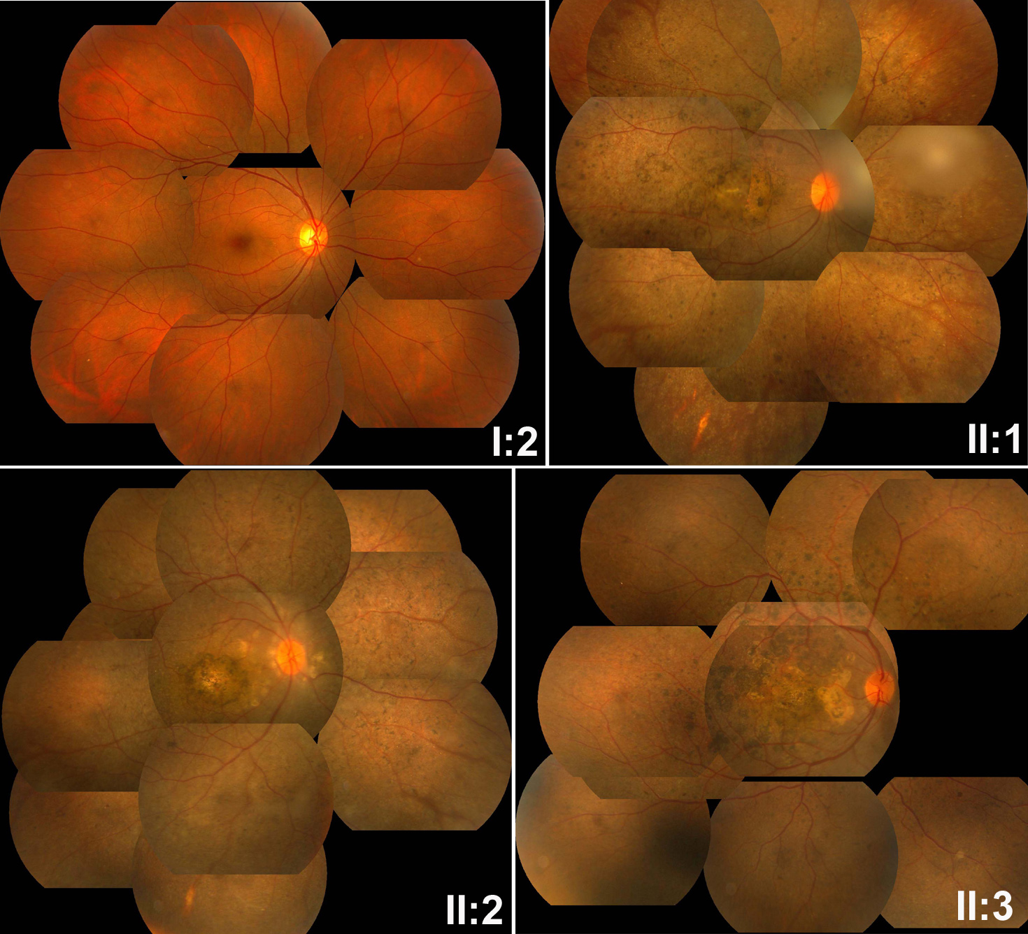
 Figure 4 of
Li, Mol Vis 2011; 17:3326-3332.
Figure 4 of
Li, Mol Vis 2011; 17:3326-3332.