Figure 4. Electron microscopy showed
apoptosis in RGC-5 cells after stable transfection of
sihoptineurin-3. A: Nuclear heterochromatin margination,
partial membrane dissolution, rough endoplasmic reticulum
expansion, and mitochondrial reduction were observed in
sihoptineurin-3 RGC-5 cells. B: Mitochondrial outer
membrane damage, cell membrane partial dissolution, and
apoptotic bodies were observed in sihoptineurin-3 RGC-5 cells. C,
D: In sihoptineurin-NC RGC-5 and blank cells, we observed
nuclear membrane integrity, with organelles enriched in
cytoplasm.
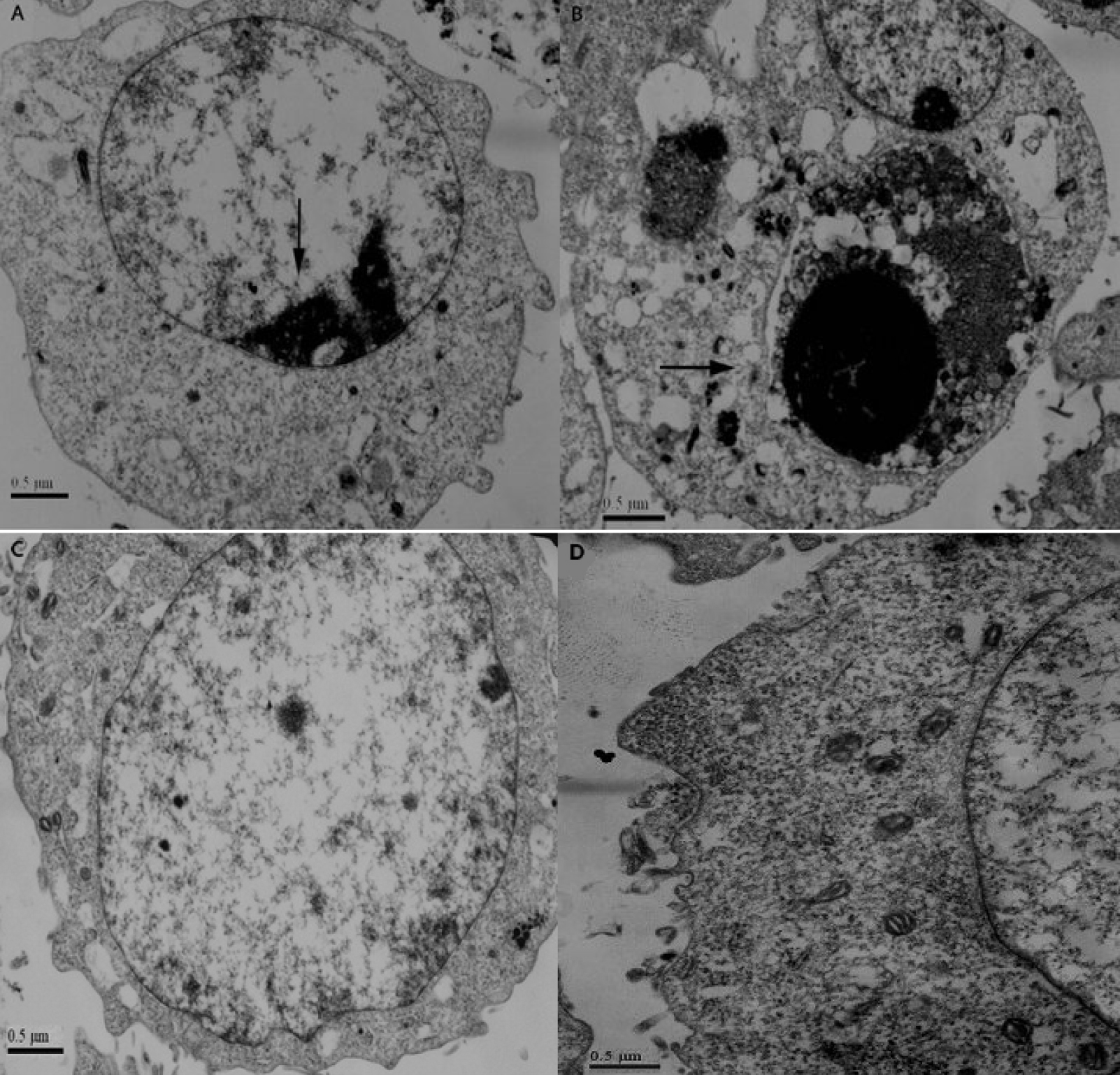
 Figure 4
of Li, Mol Vis 2011; 17:3314-3325.
Figure 4
of Li, Mol Vis 2011; 17:3314-3325.