Figure 5. Light exposure causes
massive calcium influx in RGC-5 cells. A-D: Calcium
influx was detected with fura-2 with an inverted fluorescence
microscope. A, B: Control; C, D: Cells treated
with 2,600 lx light for 2 days; Scale bar=30 μm. E:
Expanded overlay of typical responses was determined with
calcium indicator. RGC-5 cells were cultured under the 2,600 lx
light or with no light for 2 or 3 days. The intracellular free
calcium concentration was measured using the fura-2/AM
methodology. [Ca2+]i was significantly increased by
light exposure, as indicated by the green fluorescence. F:
RGC-5 cells were pre-cultured in normal media containing cobalt
(50–200 μmol/l) and then exposed to 2,600 lx light for 3 days.
Cell viability was determined with the MTT assay. Pre-treatment
with 50–200 μmol/l cobalt significantly increased cell viability
after light exposure. The results are expressed as a percentage
of the control cells and are mean values±SEM (n=48, three
separate cultures). (p<0.001, one-way ANOVA and Bonferroni
test).
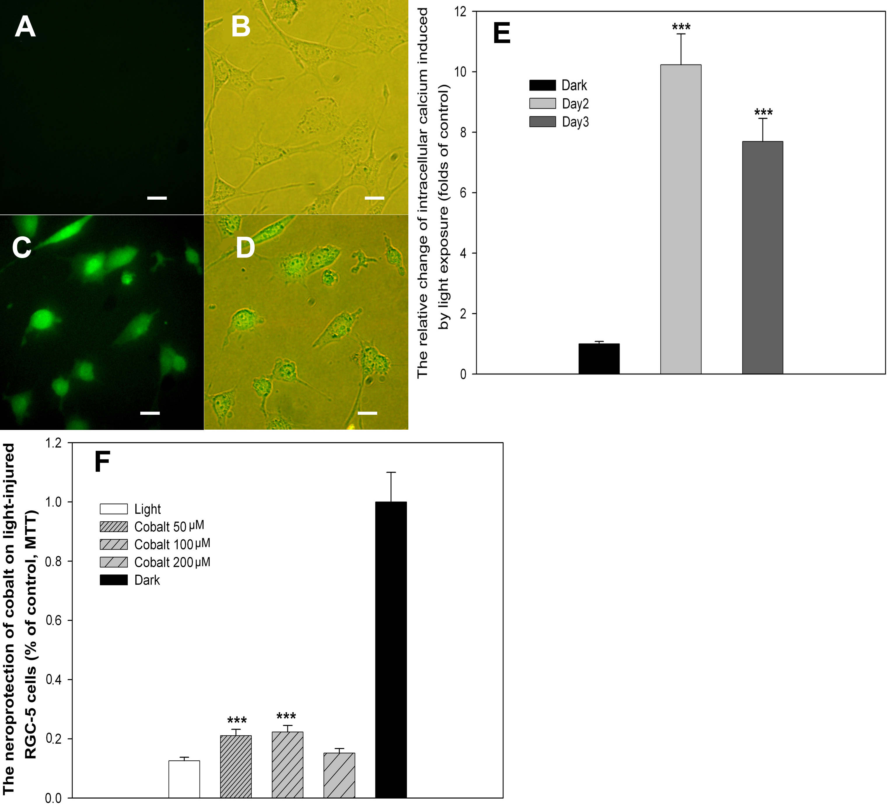
 Figure 5
of Li, Mol Vis 2011; 17:3279-3289.
Figure 5
of Li, Mol Vis 2011; 17:3279-3289.