Figure 4. PARG and AIF inhibitors
partially protect RGC-5 cells. A: The neuroprotective
effect of a PARG inhibitor on light-damaged RGC-5 cells. RGC-5
cells were pretreated with the PARG inhibitor TA and then
exposed to 2,600 lx light for 3 days. Cell viability was
determined with the MTT assay. Treatment with TA significantly
increased cell viability after light exposure. The results are
expressed as a percentage of the control cells and are mean
values±SEM (n=48, three separate cultures) (***p<0.001,
one-way ANOVA and Bonferroni test). B-G: Following light
exposure (2,600 lx, 3 days), some cells show AIF localization in
the nucleus (*) while AIF in the control cells is primarily in
the cytosol (arrows). B, C, D: dark
control; E, F, G: light exposure. Scale
bar=15 μm. H: Adding the AIF inhibitor NP (10 μM) to the
culture medium attenuates the detrimental effect of light (2,600
lx for 3 days) on RGC-5 cells. Cell viability was measured with
the MTT assay. Results are mean values±SEM for 3 different
cultures. Each result was analyzed in quadruplet. Significant
differences (***p<0.001) were determined with one-way ANOVA
and Bonferroni tests.
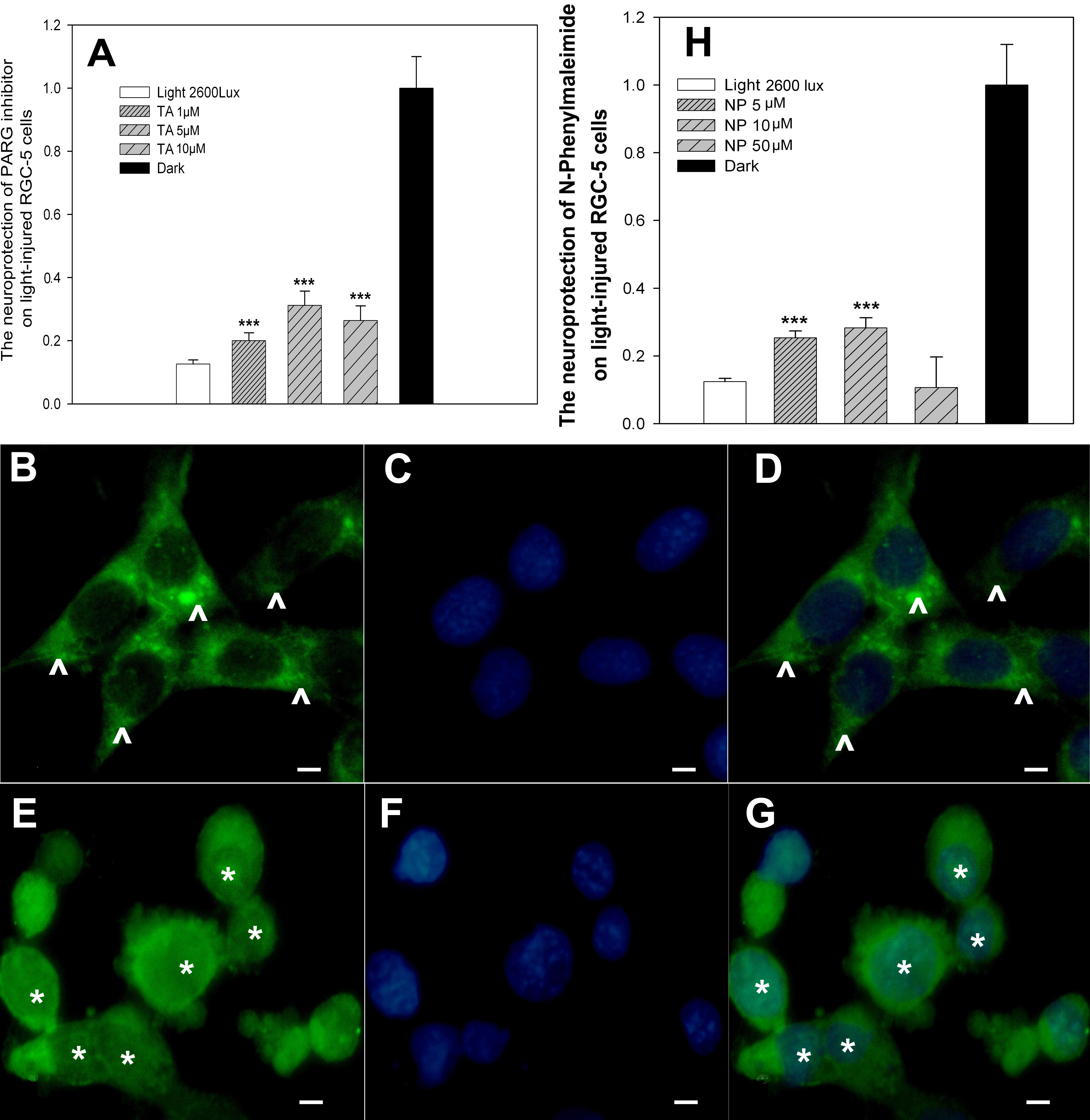
 Figure 4
of Li, Mol Vis 2011; 17:3279-3289.
Figure 4
of Li, Mol Vis 2011; 17:3279-3289.