Figure 2. Fundus photos of three
patients and a normal control. II:2 and II:7 demonstrated the
typical macular atrophy observed in the six adult patients.
III:1 at two and half years old showed mild granular retinal
pigment epithelial changes, with the tiny yellowish-white
deposits in the macula observed in the two youngest patients.
Temporal pallor of the optic disc was observed in patients II:2,
II:7, and III:1. Mild artery attenuation was noticed in II:2 and
II:7. Ctr: fundus photo of a normal control.
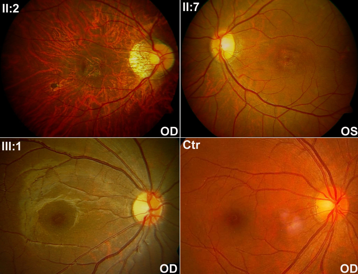
 Figure 2
of Xiao, Mol Vis 2011; 17:3271-3278.
Figure 2
of Xiao, Mol Vis 2011; 17:3271-3278.