Figure 5. Ocular phenotypic
characteristics of case II 1 (only left eye details are shown) A:
Fundus photograph showing normal pigmentation and dull foveal
reflex. B: Fundus autofluorescence image of the
posterior pole revealed normal autofluorescence. C:
Spectral domain optical coherence tomography of the left eye
revealed normal foveal contour and central retinal thickness.
All relevant outer and inner retinal layers have been labeled:
the outer nuclear layer (ONL), the outer plexiform layer (OPL),
the inner nuclear layer (INL), the inner plexiform layer (IPL),
and the ganglion cell layer (GCL).
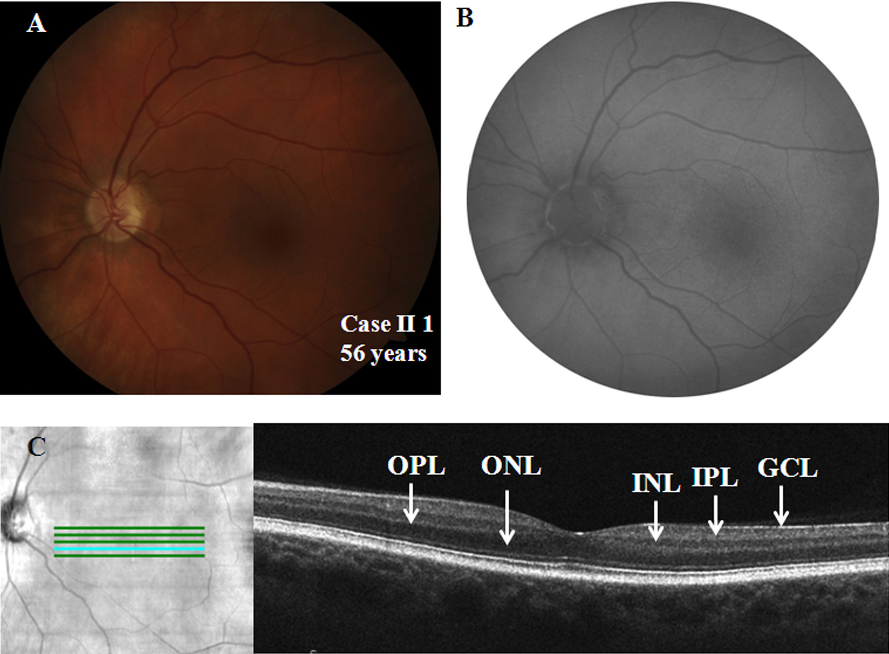
 Figure 5
of Vincent, Mol Vis 2011; 17:3262-3270.
Figure 5
of Vincent, Mol Vis 2011; 17:3262-3270.