Figure 2. Ocular phenotypic characteristics of the proband (case IV 1; only right eye details shown). A, B: Fundus photograph and red free image showing hypopigmented fundus and foveal hypoplasia, respectively. C: Spectral domain optical coherence tomography demonstrating widening of the outer nuclear layer (ONL) at the fovea. The layers
inner to ONL, including the outer plexiform layer (OPL), the inner nuclear layer (INL), and the inner plexiform layer (IPL),
are all present at the fovea consistent with foveal hypoplasia. The ganglion cell layer (GCL) is also labeled. D: Fundus autofluorescence image of the posterior pole revealed normal autofluorescence.
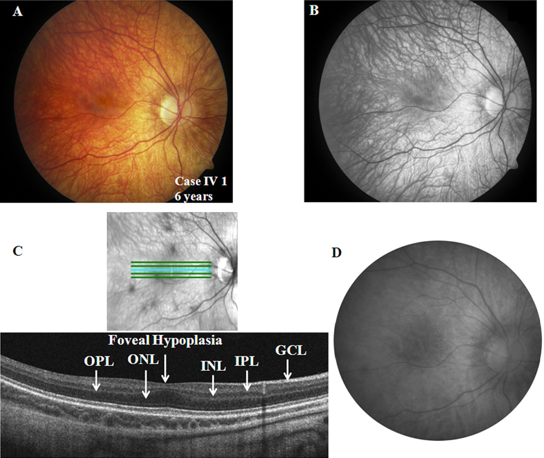
 Figure 2 of
Vincent, Mol Vis 2011; 17:3262-3270.
Figure 2 of
Vincent, Mol Vis 2011; 17:3262-3270.