Figure 4. Arginine vasopressin
secretion was not detected by stimulation with high potassium
solution. The arginine vasopressin (AVP) concentration was
assayed at 0 min (before stimulation), 15 min, and 30 min after
the retina pieces were incubated in the control solution or the
high potassium (50 mM) solution (see Methods). The concentration
of AVP at each time was estimated under the condition that the
concentration was set to 0 in the solution before incubation of
the retina. The delta (Δ) AVP concentration shown here is
defined as the difference between the concentration of AVP each
time and the concentration before the incubation. No significant
differences were found (one-way ANOVA with Tukey’s post hoc
test). Error bars indicate standard error of the mean (SEM).
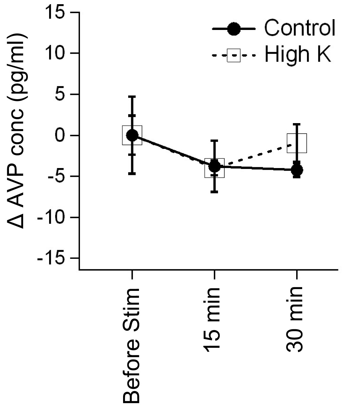
 Figure 4
of Moritoh, Mol Vis 2011; 17:3254-3261.
Figure 4
of Moritoh, Mol Vis 2011; 17:3254-3261.