Figure 1. Endogenous arginine
vasopressin–positive cells in the arginine vasopressin–eGFP
transgenic rat retina. Arginine vasopressin (AVP)/eGFP-positive
cells in (A-C) the ganglion cell layer (GCL) and (D-F)
inner nuclear layer (INL) of the AVP-eGFP transgenic rat retina
in a whole-mount preparation. A: AVP-eGFP expression in
the GCL under a fluorescence microscope. B: A cell with
GFP in the GCL detected by a confocal microscope. C: The
GFP-positive cell in B
was also positive for AVP. D: AVP-eGFP expression in the
INL under a fluorescence microscope. E: Cells with GFP
in the INL, detected by a confocal microscope. There are two
distinct types of GFP-positive cells: brighter cells and faint
cells. Asterisks show faint cells. F: The brighter
GFP-positive cell in E was also positive for AVP. The
scale bars in A and D are 50 μm. The scale bars
in B, C, E, and F are 10 μm. G:
Density recovery profile (DRP) of GFP-positive cells in the GCL
and brighter GFP-positive cells in the INL (n=7 retina pieces).
Error bars indicate standard error of the mean (SEM).
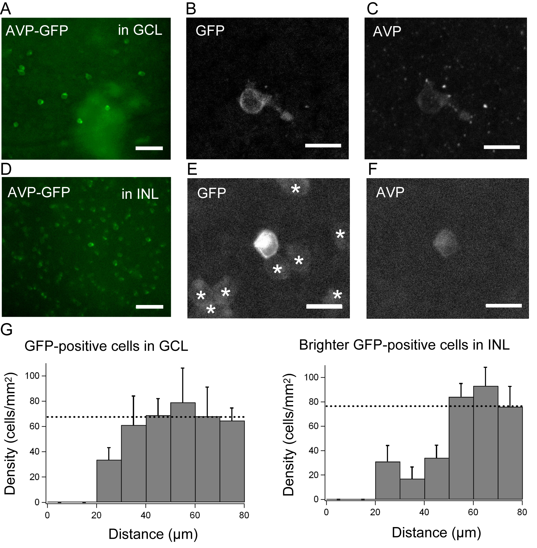
 Figure 1
of Moritoh, Mol Vis 2011; 17:3254-3261.
Figure 1
of Moritoh, Mol Vis 2011; 17:3254-3261.