Figure 4. Apoptosis profiles of HLECs at 96 h after infection by different vectors and treated with GCV at 20 µg/ml. Electron microscopy
showing subcellular structures of infected HLECs. The Lenti-IRES-EGFP infected cell nuclei were intact (A; magnification, 4880×). The enhanced specific lentiviral vector combination infected HLECs appeared as apoptotic bodies (B; magnification, 6550×). Necrosis (cell swelling, rounded mitochondrion, some subcellular structures) was seen in the cytoplasm
(C; magnification, 4880×). The percentages of apoptotic HLECs and RPECs infected by vectors: Lenti-hPGK-EGFP-HSVtk=90.12% and
89.68%, respectively; LTKCRE=53.2% and 8.62%, respectively, the enhanced specific lentiviral vector combination=87.23% and
10.12%, respectively; Lenti-IRES-EGFP=0.96% and 0.75%, respectively.
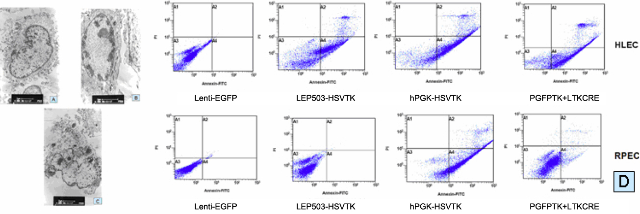
 Figure 4 of
Jiang, Mol Vis 2011; 17:291-299.
Figure 4 of
Jiang, Mol Vis 2011; 17:291-299.