Figure 6. Wild-type rhodopsin
quantification and localization in heterozygous ninaE
mutants expressing rhodopsin with a 1D4 monoclonal antibody tag
(Rh1-ID4). A: Lysates equivalent of one head were loaded
onto each lane and probed with anti-Rh1–1D4 antibody that
specifically recognizes wild-type protein. Arrestin2 was used as
the loading control. B: The blot probed with
anti-Rh1–1D4 antibody was quantified to show levels of rhodopsin
in mutants relative to the wild type. The reduction in levels of
soluble wild-type rhodopsin in heterozygous mutant flies is
significant compared to controls (p<0.0001). C-E:
Dark-reared fly retinas were isolated and stained for F-actin
and Rh1–1D4. Wild-type photoreceptors show proper rhodopsin
localization to the base of the rhabdomeres. F-H:
Flies expressing non-1D4-tagged rhodopsin (+/+) show no staining
for rhodopsin, highlighting the specificity of the antibody. I-K:
Flies haploid for mutant ninaER11 and
wild-type Rh1–1D4 show partial proper rhodopsin localization
with significant rhodopsin-positive puncta mislocalized to the
cell body. Error bars are represented as average % rhodopsin±SD.
Each column represents the average of three biologic replicates.
Scale bar represents 5 μm.
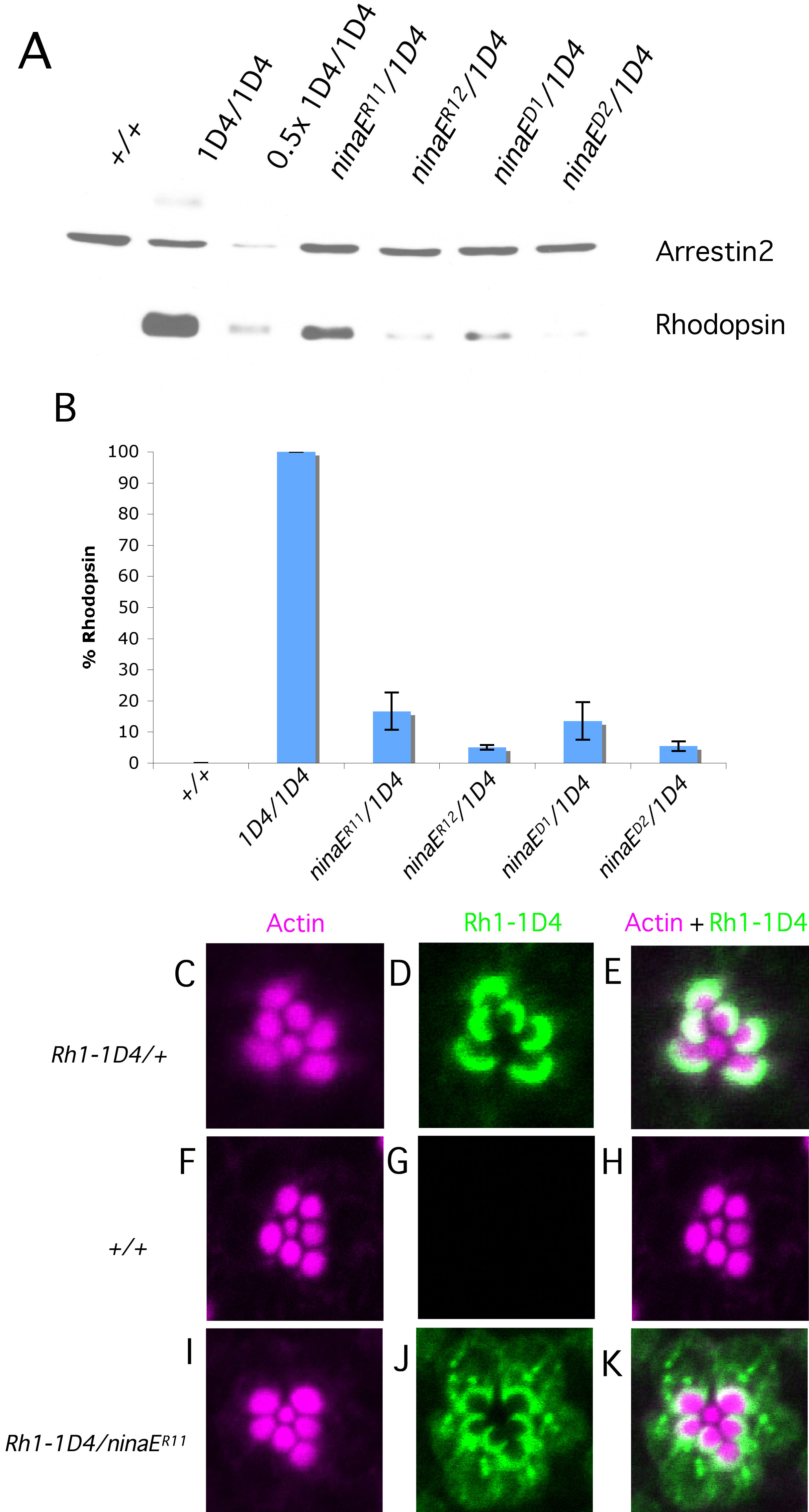
 Figure 6
of Mitra, Mol Vis 2011; 17:3224-3233.
Figure 6
of Mitra, Mol Vis 2011; 17:3224-3233.