Figure 5. Quantification of soluble
and total rhodopsin in heterozygous ninaE mutants. A:
Lysates equivalent of four heads were loaded onto a sodium
dodecyl sulfate PAGE (SDS–PAGE) gel and probed with anti-Rh1
antibody, while Rab7 antibody was used as a loading control for
western analysis. B: Whole head lysates equivalent to
two wild-type heads and four heterozygous mutant heads were
prepared and diluted for slot blot analysis as detailed in the
Methods and probed with anti-Rh1 antibody. C: The blots
probed with anti-Rh1 antibody were quantified to reflect the
relative level of soluble versus total rhodopsin in mutant
flies. Note the loss of wild-type rhodopsin intensity in (C)
when mutant rhodopsin is co-expressed in the same
photoreceptors. The difference in the levels of soluble versus
total rhodopsin in each of the genotypes tested is statistically
significant (p<0.014). Error bars are represented as average
% Rhodopsin±SD. Each column represents the average of three
biologic replicates.
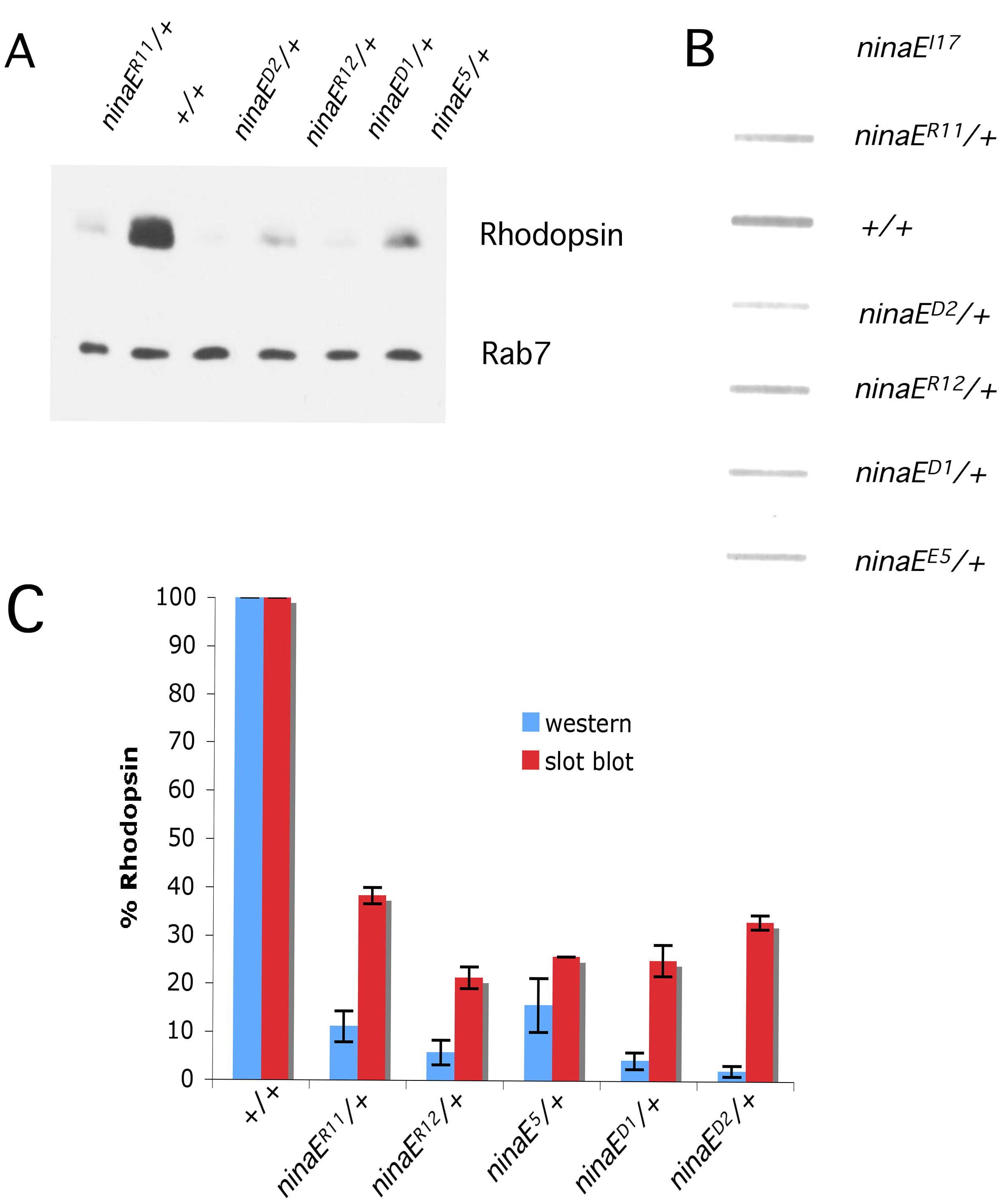
 Figure 5
of Mitra, Mol Vis 2011; 17:3224-3233.
Figure 5
of Mitra, Mol Vis 2011; 17:3224-3233.