Figure 4. Rhodopsin and NinaA show
improper localization in heterozygous ninaE mutants.
Dark-reared fly retinas were isolated and triple stained for
F-actin, Rhodopsin (Rh1), and neither inactivation nor
afterpotential protein (NinaA). A-D: Wild-type
photoreceptors show proper rhodopsin localization to the base of
the rhabdomeres and NinaA is localized to the cell body. E-H:
ninaER11/+ photoreceptors show partial proper
rhodopsin localization with multiple rhodopsin- and
ninaA-positive puncta present in the cell body. NinaA staining
is restricted to the edges of the cell body. I-L:
ninaER12/+ photoreceptors show rhodopsin
localization to the base of the rhabdomeres, with diffuse
rhodopsin staining in the cell body having few distinct
rhodopsin-positive vesicles. NinaA stains diffusely throughout
the cell body similar to the wild type. Scale bar represents 5
μm.
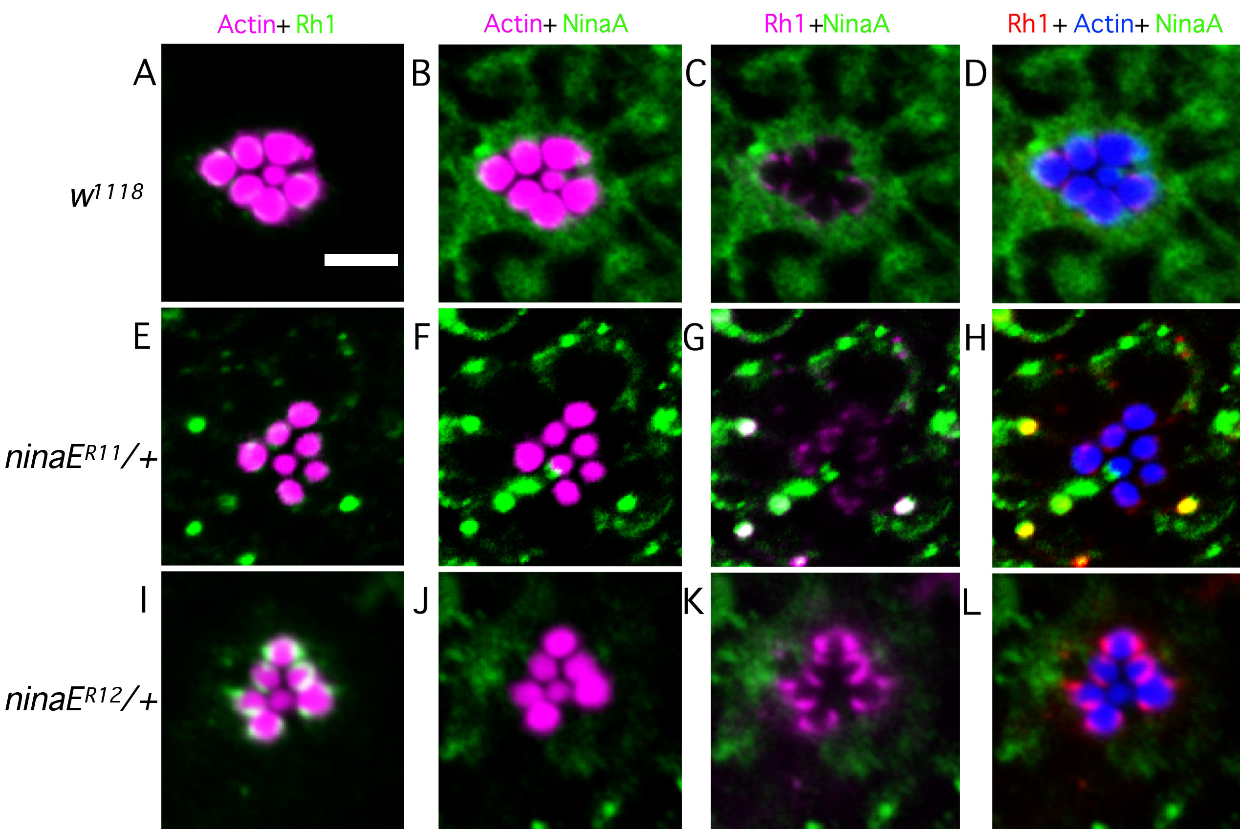
 Figure 4
of Mitra, Mol Vis 2011; 17:3224-3233.
Figure 4
of Mitra, Mol Vis 2011; 17:3224-3233.