Figure 3. Effects of mapracorat and
dexamethasone on cytokine-sustained eosinophil survival. Control
eosinophils were routinely cultured in the presence of 10% fetal
bovine serum or for 48 h in 0.1% fetal bovine serum and treated
with granulocyte-macrophage colony stimulating factor (70 pM; A)
or interleukin-5 (30 pM; B) added concomitantly with
mapracorat or dexamethasone (0.001–10 μM) or their vehicle.
Apoptosis was determined by flow cytometry, evaluating the
cell’s ability to bind annexin V and exclude propidium iodide as
described under Methods. Results are expressed as percentages of
apoptotic cells. Data are presented as mean±standard error of
the mean from six experiments performed in triplicate using
different eosinophil cell cultures. **Versus the respective
control; p value (p)<0.01. ##Versus 0.1% fetal
bovine serum; p<0.01 §§Versus 0.1% fetal bovine
serum+granulocyte macrophage-colony stimulating factor or versus
0.1% fetal bovine serum + interleukin-5; p<0.01.
Abbreviations: Ctrl represents controls; FBS represents fetal
bovine serum; GM-CSF represents granulocyte-macrophage colony
stimulating factor; IL-5 represents interleukin-5.
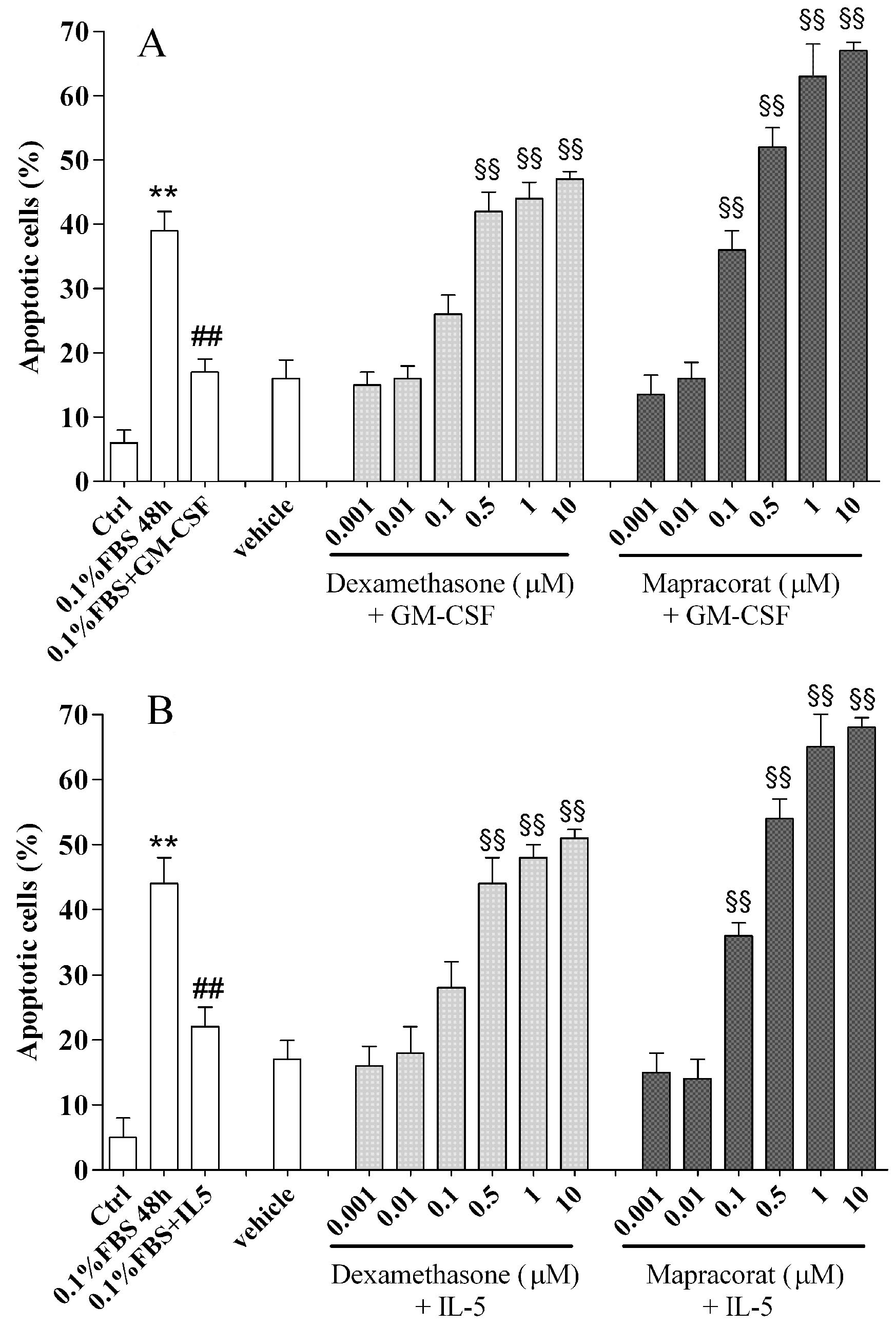
 Figure 3
of Baiula, Mol Vis 2011; 17:3208-3223.
Figure 3
of Baiula, Mol Vis 2011; 17:3208-3223.