Figure 2. Mapracorat and
dexamethasone induce caspase-3 activation in human eosinophils.
Control cells were cultured for 24 h in 0.1% fetal bovine serum
and in the absence of granulocyte-macrophage colony stimulating
factor and interleukin-5 and were treated with mapracorat or
dexamethasone (0.001–10 μM). Alternatively, eosinophils were
exposed to the vehicle alone. A: A representative
western blot, repeated at least six times using different
eosinophil cell cultures, with similar results, showing the
bands of apparent molecular weights of caspase-3 of
approximately 32 kDa and 17 kDa and beta-actin of
approximately 42 kDa. B: Densitometric analysis of
the bands of caspase-3 fragment of approximately 32 kDa. C:
Densitometric analysis of the bands of caspase-3 fragment of
approximately 17 kDa. The approximate molecular mass of the
fragments of 32 and 17 kDa was determined by comparison
with molecular mass standards. The relative optical density of
each band was determined by densitometry and defined by
normalization of the bands of capsase-3 32 kDa or
17 kDa to the β-actin band. A total of 50 μg of protein
extract was loaded and separated in a polyacrylamide gel, as
described under “Methods..” Data are presented as mean±standard,
n=6. *Versus controls; p value (p)<0.05. **Versus controls;
p<0.01. ***Versus controls; p<0.001. Abbreviations: Ctrl
represents controls; p32 represents apparent molecular weight of
caspase-3 of approximately 32 kDa; p17 represents apparent
molecular weight of caspase-3 of approximately 17 kDa.
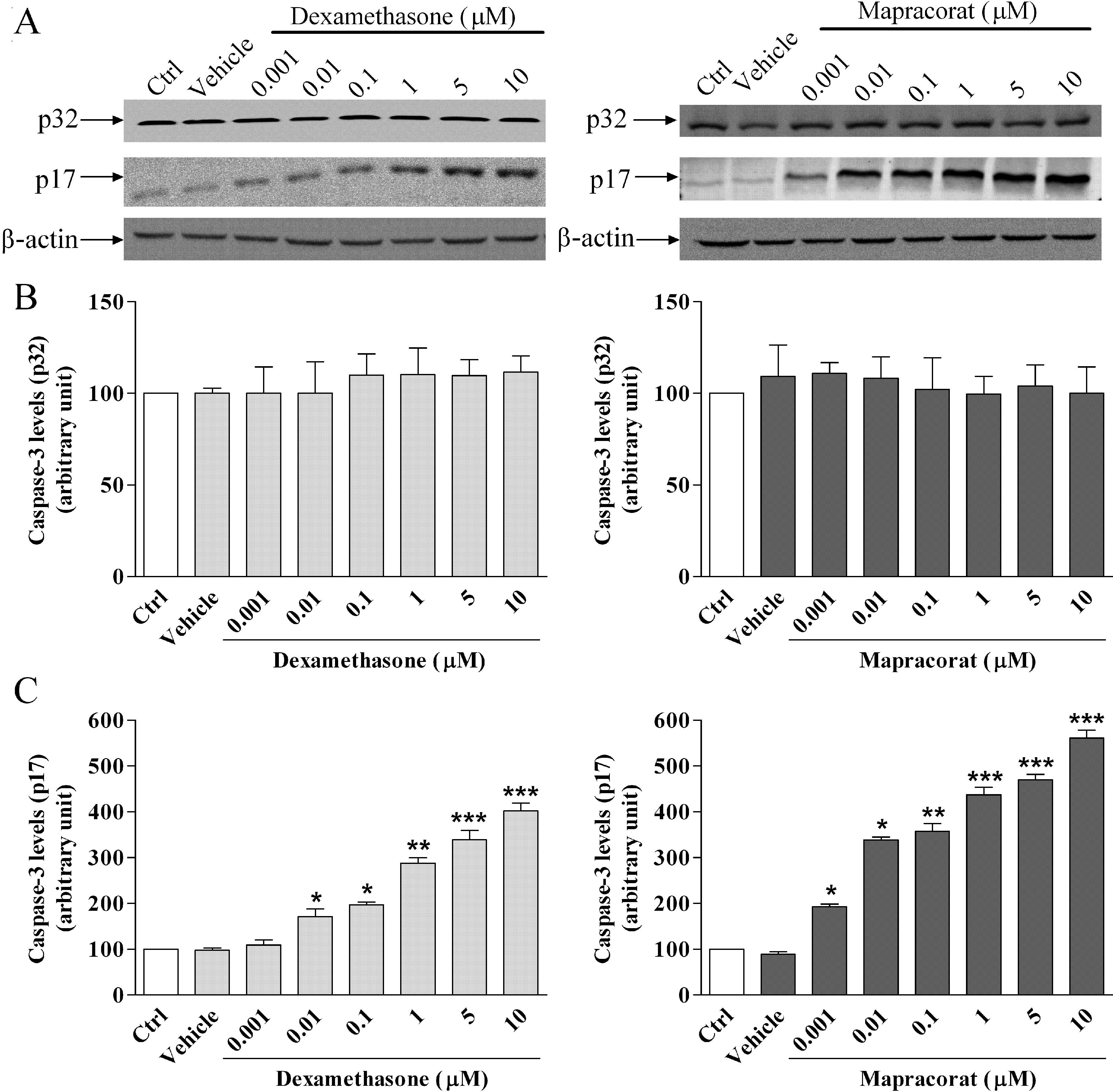
 Figure 2
of Baiula, Mol Vis 2011; 17:3208-3223.
Figure 2
of Baiula, Mol Vis 2011; 17:3208-3223.