Figure 1. Effects of mapracorat and
dexamethasone on spontaneous eosinophil apoptosis. A:
Peripheral human blood eosinophils cultured up to 48 h in 0.1%
fetal bovine serum (FBS) and in the absence of
granulocyte-macrophage colony stimulating factor and
interleukin-5 show time-dependent apoptosis. Mapracorat and
dexamethasone (0.001–10 μM) or their vehicle were added for 48
h. Control cells (Ctrl) were cultured in RPMI+10% FBS.
Mifepristone (10 μM) was co-incubated with mapracorat or
dexamethasone (5 or 10 μM). Apoptosis was determined by flow
cytometry, evaluating the cell’s ability to bind annexin V and
exclude propidium iodide as described under Methods. Results are
expressed as percentages of apoptotic cells. Data are presented
as mean±standard error of the mean from six experiments
performed in triplicate using different eosinophil cell
cultures. *Versus controls; p value (p)<0.05. **Versus
controls; p<0.01. #Versus vehicle; p<0.01. ##Versus
mapracorat or dexamethasone 5 or 10 μM; p<0.01. §Versus
dexamethasone at the same concentration; p<0.01. B: A
representative experiment showing total percentage of apoptotic
eosinophils (annexin V+/propidium iodide- and annexin
V+/propidium iodide+ cells). Abbreviations: Ctrl
represents controls; 0.1 FBS represents 0.1% fetal bovine serum;
MF represents mifepristone; Dex represents dexamethasone; Map
represents mapracorat.
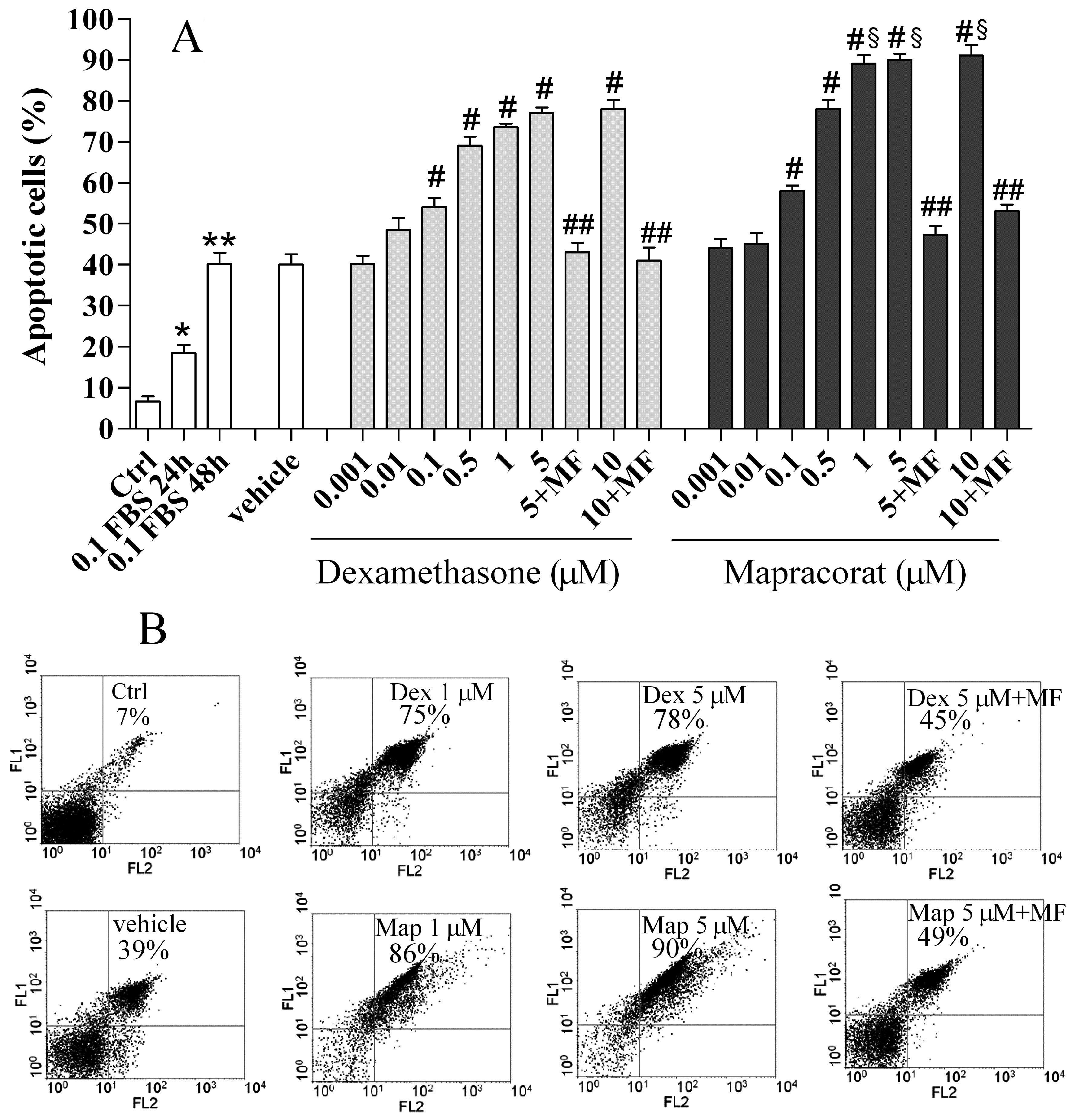
 Figure 1
of Baiula, Mol Vis 2011; 17:3208-3223.
Figure 1
of Baiula, Mol Vis 2011; 17:3208-3223.