Figure 2. Slit-lamp photographs showing granular deposits distributed in the left eye (A, individual III:13; B, individual III:15). The arrow indicates liner opacities in the superficial stroma. Confocal images showing numerous hyper-reflective
dots with sharp shapes scattered between stromal cells and nerve fibers in the superficial corneal stroma in the left (C) and right eye (D) both of individual III:13.
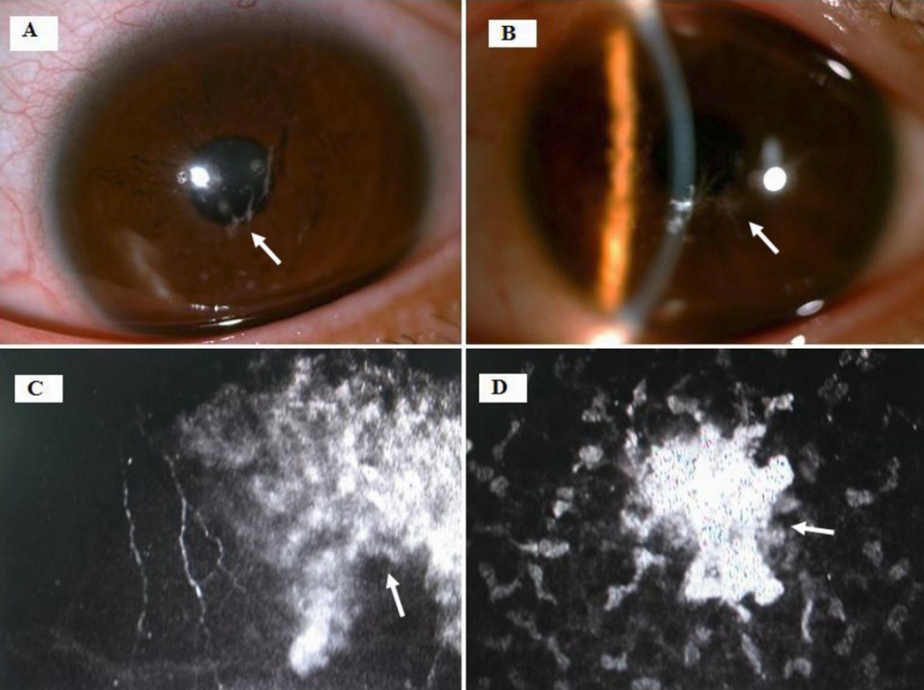
 Figure 2 of
Gu, Mol Vis 2011; 17:3200-3207.
Figure 2 of
Gu, Mol Vis 2011; 17:3200-3207.