Figure 6. Development of acellular capillaries in the diabetic retina. A-D: Representative retinal trypsin digest (RTD) images showing the effect of fibronectin (FN)-siRNA on the development of acellular
capillaries (as indicated by arrows) in retinas of a (A) normal rat, (B) diabetic rat, (C) diabetic rat intravitreally injected with FN-siRNA, and (D) diabetic rat intravitreally injected with scrambled siRNA. FN-siRNA treatment in the diabetic rats specifically reduced
the development of acellular capillaries compared to those of uninjected diabetic rats. E: Graphical representation of acellular capillaries in four groups of rats: normal, diabetic, diabetic injected with FN-siRNA,
and diabetic injected with scrambled siRNA, showing that an intravitreal injection of FN-siRNA reduces the development of
acellular capillaries in diabetic rat retinas. Normal versus Diabetic *=p<0.05; Diabetic versus Diabetic+FN-siRNA **=p<0.05,
n=8.
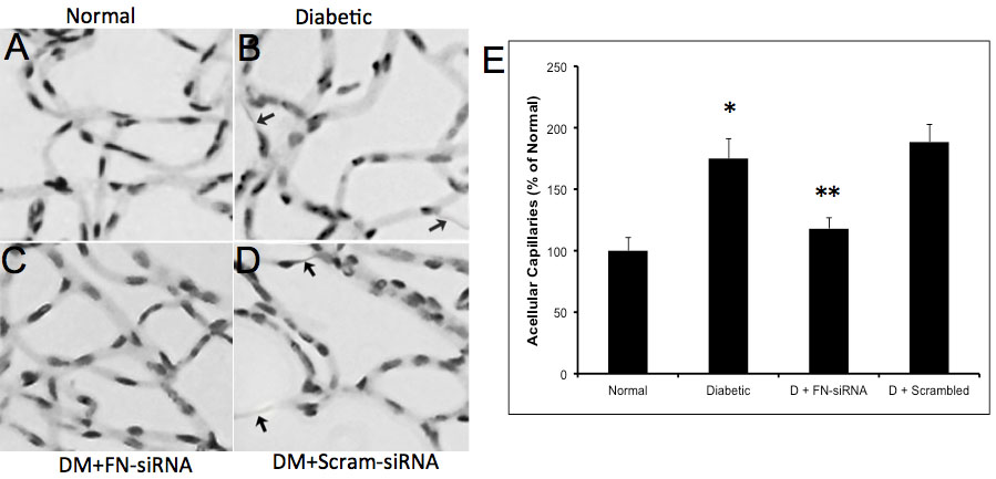
 Figure 6 of
Roy, Mol Vis 2011; 17:3166-3174.
Figure 6 of
Roy, Mol Vis 2011; 17:3166-3174.