Figure 5. Changes in MAPK activation
patterns and IL-6/8 release in DUSP1 subline. A: Western
blot analysis of total DUSP1 protein expression in resting
scrambled HCEC and DUSP1 subline. B: Comparison of time
dependent changes in MAPK phosphorylation in scrambled shRNA and
DUSP1subline. Cells were exposed to CAP (20 µM) for indicated
times. Changes are compared in p-ERK1/2, p-JNK1/2, and p38 MAPK
in two different sublines. Protein loading equivalence validated
based on invariant ERK1/2 expression levels. C: DUSP1
gene knockdown enhances CAP-induced IL-6 and IL-8 release. ELISA
was performed on scrambled shRNA and DUSP1 subline after 24 h
exposure to CAP (20 µM). Three independent experiments each
performed in triplicate.
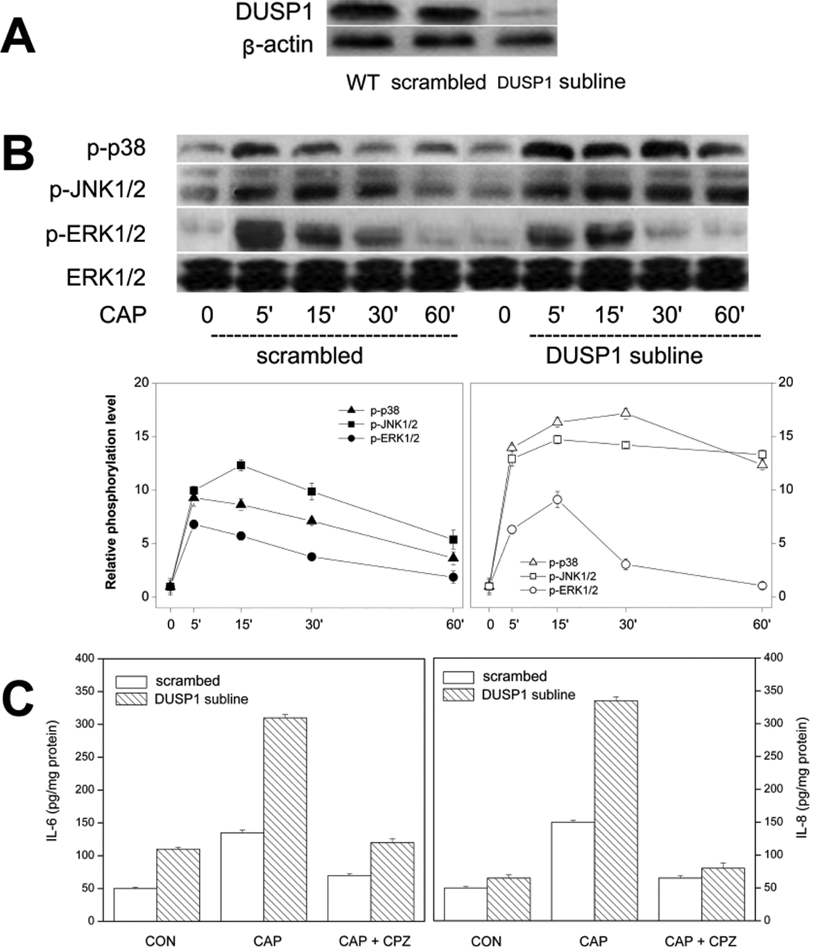
 Figure 5
of Wang, Mol Vis 2011; 17:3137-3146.
Figure 5
of Wang, Mol Vis 2011; 17:3137-3146.