Figure 4. A selection of transmission
electron micrographs from the corneal stroma of guinea pigs fed
a normal (AA+) or ascorbic acid deficient (AA-) diet, with
(UVB+) or without (UVB-) daily UV-B exposure. No clear
differences in collagen fibril organization were evident between
treatment groups and a disruption of cellular organelles was
seen in all corneas (A-D). Regions devoid of
regularly arranged collagen fibrils (arrows) were particularly
evident in the UVB-treated corneas (C and D).
Original magnification: 3000× (A, C, D)
and 5000× (B).
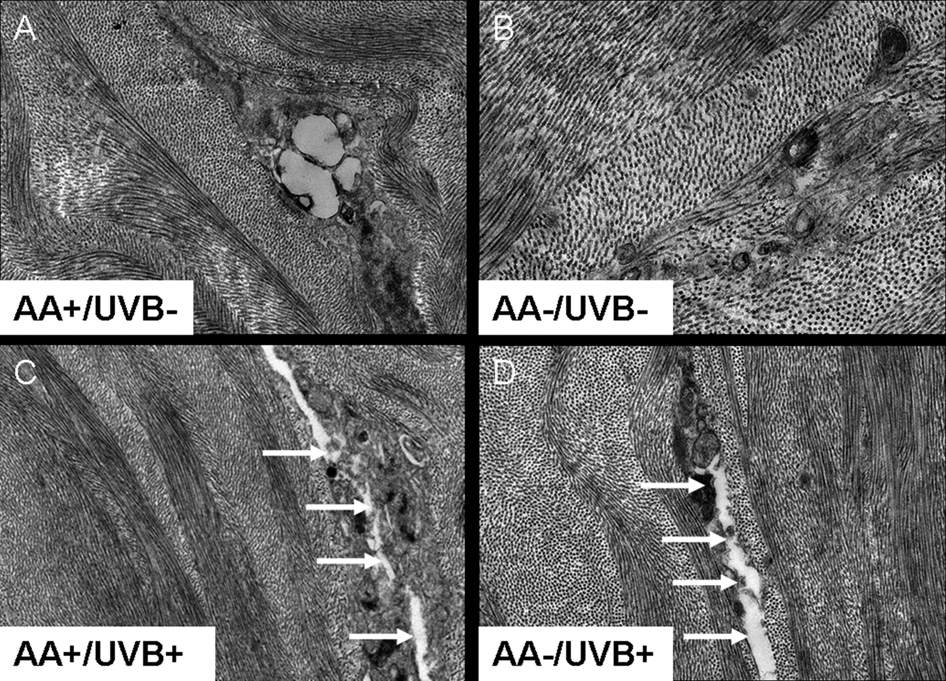
 Figure 4
of Hayes, Mol Vis 2011; 17:3107-3115.
Figure 4
of Hayes, Mol Vis 2011; 17:3107-3115.