Figure 5. Fundus photographs and optical coherence tomography (OCT) scans of patient C-4. A, B: Bilateral vitelliform discs in the vitelliruptive stage. C, D: Bilateral hyperreflective dome-shaped lesions located between the RPE and the neuroretina. OD represents the right eye;
OS represents the left eye.
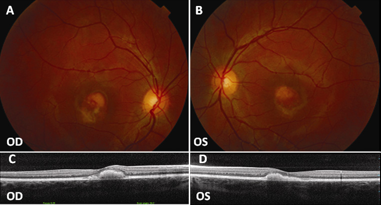
 Figure 5 of
Sodi, Mol Vis 2011; 17:3078-3087.
Figure 5 of
Sodi, Mol Vis 2011; 17:3078-3087.