Figure 4. Fundus photographs and optical coherence tomography (OCT) scans of patient C-3. A, B: Bilateral vitelliform discs in the vitelliruptive stage with retinal pigment epithelium (RPE) dystrophy inferiorly to the
macula. C, D: Macular optically empty lesions containing partially reabsorbed hyperreflective material. OD represents the right eye; OS
represents the left eye.
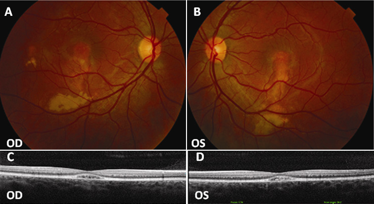
 Figure 4 of
Sodi, Mol Vis 2011; 17:3078-3087.
Figure 4 of
Sodi, Mol Vis 2011; 17:3078-3087.