Figure 3. Fundus photographs and optical coherence tomography (OCT) scans of patient B-3. A: Normal fundus in right eye (OD) and (B) vitelliform disc in the vitelliruptive stage in left eye (OS). C: Normal macular profile in OD and (D) central RPE detachment with hyperreflective material in OS.
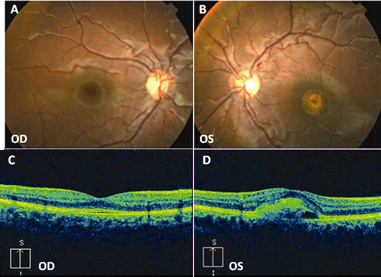
 Figure 3 of
Sodi, Mol Vis 2011; 17:3078-3087.
Figure 3 of
Sodi, Mol Vis 2011; 17:3078-3087.