Figure 2. Fundus photographs, fluorescein angiography, and optical coherence tomography (OCT) scans of patient A-3. A, B: Bilateral vitelliform disc in the vitelliruptive stage resulting in (C, D) a macular hypofluorescent lesion with irregular areas of hyperfluorescence and (B) smaller multifocal vitelliform discs resulting in (D) hypofluorescent areas in left eye (OS). E, F: Bilateral macular optically empty lesions with clumping of hyperreflective material on the posterior retinal surface and
some irregular thickening of the RPE layer. OD represents the right eye.
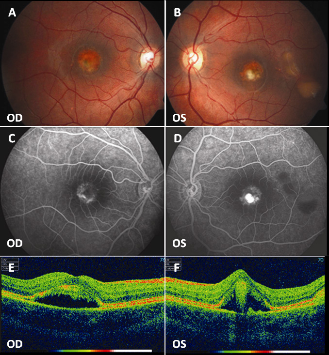
 Figure 2 of
Sodi, Mol Vis 2011; 17:3078-3087.
Figure 2 of
Sodi, Mol Vis 2011; 17:3078-3087.