Figure 1. Effects of IL-17A on phosphorylation of Erk1/2, p38MAPK, or Akt in ARPE-19 cells. ARPE-19 cells were incubated without or
with 100 ng/ml IL-17A whereafter the degree of phosphorylation of Erk1/2, p38 and Akt was measured. The amounts of intracellular
phosphorylated signaling molecules in 10,000 permeabilized ARPE-19 cells were measured by flow cytometry. A representative
example and MFI result expressed as the mean±SD of three independent experiments are shown for the phosphorylation of Erk1/2
(20 min stimulation; A), p38 MAPK (20 min stimulation; B), and Akt (20 min stimulation; C) in response to IL-17A (thick line histograms). Thin line histograms indicate basal phosphorylation in the absence of IL-17A.
Western blot analysis of phosphorylated Erk1/2, p38MAPK or Akt was performed on ARPE-19 cells stimulated with or without 100
ng/ml of IL-17A for the indicated time periods. Results are representative of three separate experiments.
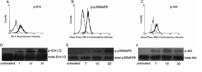
 Figure 1 of
Chen, Mol Vis 2011; 17:3072-3077.
Figure 1 of
Chen, Mol Vis 2011; 17:3072-3077.