Figure 4. Characterization of lop11
lenses. The persistence of nuclei in lop11 lens fiber
cells (arrows) was first observed in P0.5 lenses without any
other significant morphological differences noted between wild
type (A, B) and lop11 (C, D) lenses; the
same findings were noted in P7 wild type (E, F) and lop11
(G, H) lenses. Quantification of lens fiber nuclei number
in the lens cross-sections in P0.5 and P7.5 wild type and lop11
lenses is shown in K. The error bars represent the SEM
and asterisks indicate significant differences (p<0.05; t-test)
calculated from comparison with wild-type. Immunohistochemistry
of P0.5 wild type (I) and lop11 (J) lenses
using anti-MIP antibody showing lens fiber cell staining. L:
BrdU incorporation in the lens epithelial cells of E16.5 mice.
Percentages of BrdU-positive cells are shown from six different
animals. Error bars represent SEM. The scale bars in A,
C, E, and G indicate 250 µm, whereas B,
D, F, and H are 50 µm. All sections are
cut in the center of the lens, containing the pupil and optic
nerve.
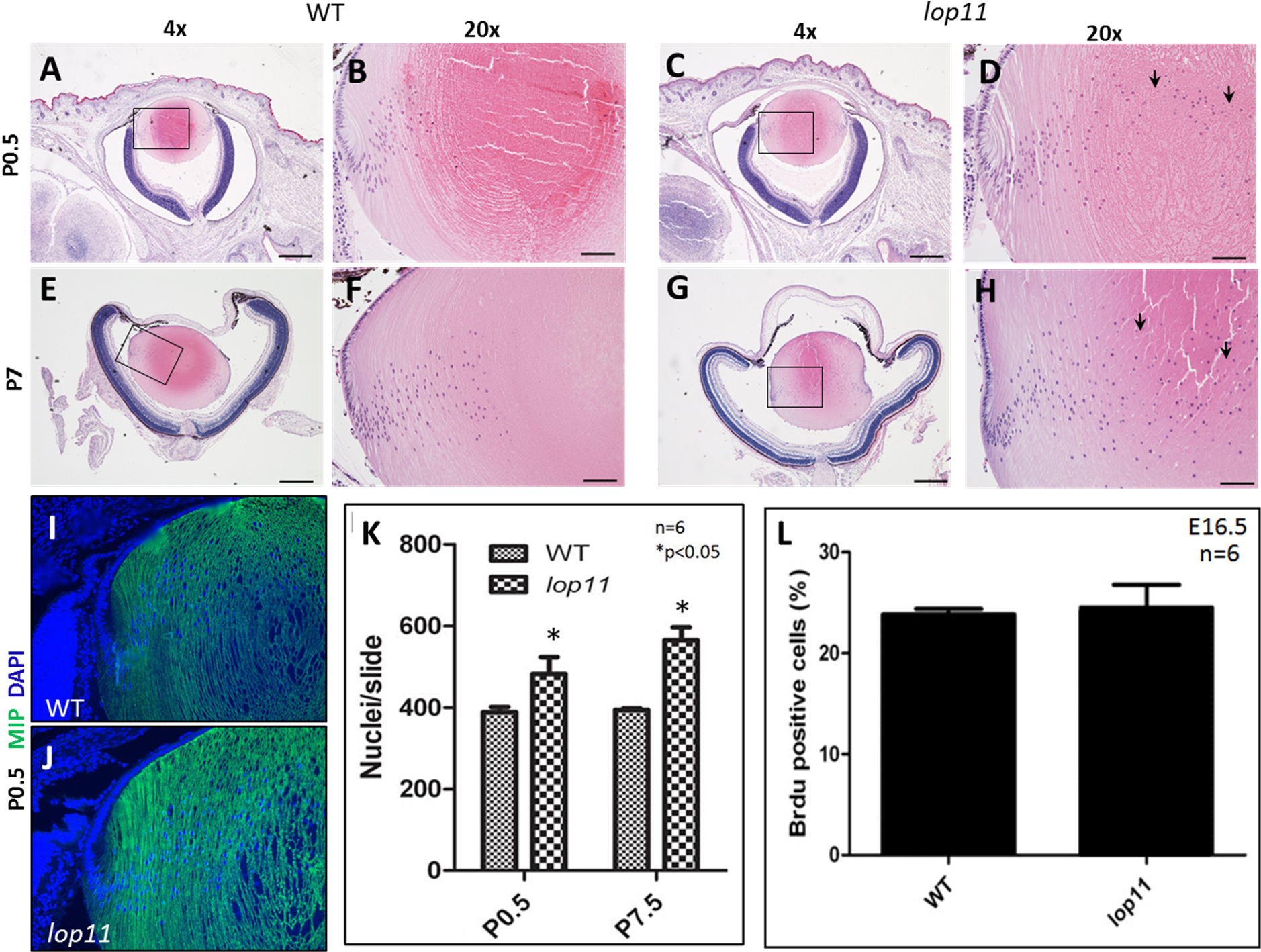
 Figure 4
of Liang, Mol Vis 2011; 17:3062-3071.
Figure 4
of Liang, Mol Vis 2011; 17:3062-3071.